
Sage — Cardiology: Mitochondrial Energy → Cardiovascular Phenotype
1. Framing — compensatory cascade → cardiovascular phenotype
Chronic diseases of modernity share a recurring upstream pattern in the EnSurf map: persistent cellular bioenergetic insufficiency rooted in mitochondrial dysfunction. Aging reads as progressive loss of ATP-generating capacity; cancer as fermentative escape when oxidative headroom collapses; fuel mismatch as wrong substrates degrading the membrane theater of oxidation; organ dysfunction as named syndromes that are higher-order compensations. Cardiovascular phenotypes belong in the same geometry — vessel wall, myocardium, autonomic control under orthostasis, and (as proposed bridge below) excitability/conduction that depends on ion-pump ATP.
The standing rule (Compensatory-cascade) is simple: when ATP load exceeds ATP-generating capacity, the system reverts to more primitive (lower) nests of energy production to keep generating enough ATP to maintain persistence. Acute emergency-mode can be adaptive and reversible. Chronic emergency-mode lock-in is what we call disease. Restore capacity in time and much of the cascade can wind down; fail to restore and selection hardens emergency-adapted phenotypes. Cardiology’s named syndromes — plaque, orthostatic tachycardia, hypertrophy/fibrosis, pressure elevation, rhythm instability — are readable first as clusters of that chronic mode under different tissue clocks and loads, not as freestanding villains inventing disease beside the engine.
Stay monist: Shore / Surf Judge language names orientation inside one field — not a second “mind stuff” beside mitochondria, and not a moral story in which lipids, rate, or pressure are villains independent of cellular fuel handling (Dashboard-discipline: map ≠ terrain; instruments, not a second ontology). The bioenergetics here follows Nick Lane’s work; cascade framing, Surfer/Energy Phase, and EN overlay are EnSurf’s own extensions, not Lane’s claims.
2. Atherosclerosis — ApoB and the Engine: An Orchestra of Atherogenesis
Why ApoB Is Necessary but Not Sufficient, and Why Neither Camp's Silver Bullet Is Good Management
By Val Zudans, MD
Atherosclerosis remains the leading cause of cardiovascular death worldwide. Two camps argue about it, and both have hold of something real.
The first, articulated by clinical lipidologists such as Thomas Dayspring, MD, holds that atherosclerosis is fundamentally a disorder of cholesterol retention in the arterial wall, delivered by apoB-containing lipoproteins (LDL, VLDL remnants, Lp(a)). ApoB particles act as "delivery trucks"; their cargo, once trapped by subendothelial proteoglycans, oxidizes, triggers macrophage foam-cell formation, and drives inflammation and plaque. ApoB count is the better biomarker because each particle carries one apoB molecule. Lowering apoB reduces events in RCTs, Mendelian randomization, and familial hypercholesterolemia (FH) [1–3].
The second camp, where my own clinical observation, personal metabolic work, and the EnSurf mitochondrial optimization system began, looks upstream at the cell's energy economy. Mitochondrial function, insulin sensitivity, inflammation, and circadian health decide how vessels, liver, and macrophages handle the lipid that arrives.
The two are often framed as rivals, with apoB on one side and energy on the other. The evidence fits better as an interplay. ApoB particle number and retention are necessary but not sufficient: a real atherogenic input, without which plaque does not form, but not the whole score. Metabolic and energy-flux health sets the context that decides how much harm a given apoB exposure does, and it also shapes apoB levels themselves. Risk, in plain words, is exposure (how many apoB particles, for how long) acting on a tissue whose vulnerability depends on its metabolic state. Neither half works without the other.
A better image than two camps is an orchestra of atherogenesis. ApoB particles carry the melody, and without them there is no plaque to build. But the endothelium, the arterial matrix, the redox state of the wall, the macrophages, the inflammatory response, and the cap that holds a plaque together each play their own part, and each can be atherogenic in its own right. ApoB is necessary but not sufficient. The rest of this essay follows that score through the stages of plaque-building, with the apoB part and the metabolic and wall part at each step.
That is the thesis of this essay, and it is a management thesis as much as a biological one: looking for a silver bullet in either camp, and oversimplifying to fit it, is not ideal management. The apoB-only version says lowering the number is the whole answer. The metabolic-only version says apoB doesn't matter if you are metabolically healthy. Each holds a truth and drops one. Better care lowers exposure where it is warranted, with diet and with drugs (statins and other LDL-lowering therapies included), and lowers tissue vulnerability with metabolic, mitochondrial, and circadian levers. The plan should fit the person, not one camp's rule.
What follows is offered as a hypothesis about how the two interact, not a protocol and not a reason to stop any proven therapy.
What the ApoB Camp Gets Right: Exposure Is Necessary
In the prevailing view, retention of apoB-containing lipoproteins in the subendothelium initiates disease. Proteoglycans bind particles, which then oxidize or modify. Oxidized LDL enters macrophages via scavenger receptors, forming foam cells that release cytokines, recruit monocytes, amplify inflammation, and stimulate smooth-muscle proliferation. A necrotic core and thin fibrous cap follow, increasing vulnerability. What fills a foam cell is mostly cholesteryl ester, stored in cytoplasmic lipid droplets [4, 5].
The model correctly prioritizes apoB particle number over LDL-C alone, since each atherogenic particle carries one apoB. It notes insulin resistance worsens risk by increasing hepatic VLDL and thus apoB count. Large-scale evidence—from statin, PCSK9-inhibitor, and bempedoic acid trials to genetic studies—shows dose-dependent event reduction with apoB lowering [1–3, 201]. Genetic variants that lower triglycerides (LPL) and variants in the LDL-receptor gene (LDLR) that lower LDL-C protect to a similar degree per unit of apoB lowered, which points to the particle count itself [3]. FH, a lifelong exposure caused by a variant in a single gene (most often LDLR), produces early disease, and lifelong lowering improves survival [6]. A PCSK9 inhibitor, which acts by sparing hepatic LDL receptors, reduced events in a large outcome trial [2].
I accept this plainly. ApoB exposure is necessary for atherosclerosis, and lowering it lowers risk in proportion. Any account of atherosclerosis that treats it as a mere readout is wrong. What the evidence does not show is that apoB is sufficient on its own; the stages below show where the rest of the orchestra comes in.
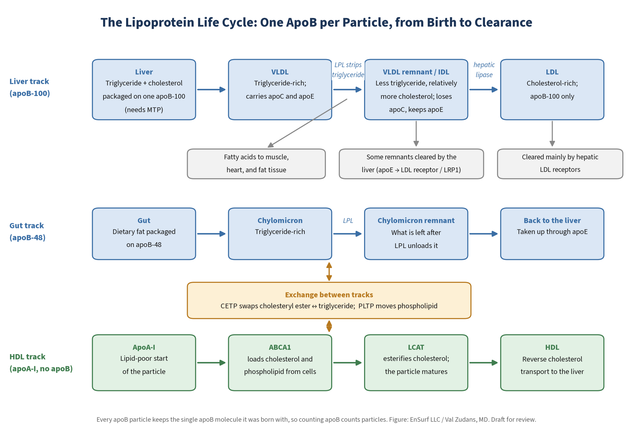
The Lipoprotein Life Cycle
Before arguing about what apoB means, it helps to follow one particle from birth to death. The whole system is a fuel-delivery network, and cholesterol rides along with the fuel.
Assembly in the Liver
The liver packages triglyceride and cholesterol around a single apoB-100 molecule, a step that requires microsomal triglyceride transfer protein (MTP) [7]. How much triglyceride the liver has to export depends on supply: fatty acids released from adipose tissue, dietary fat returning in remnants, and fat made new from carbohydrate by de novo lipogenesis (DNL) [8, 9]. Fructose is a strong driver of DNL. In a controlled feeding study, fructose but not glucose raised hepatic DNL, post-meal triglycerides, apoB, and small dense LDL [10]. A liver with more fat to export secretes more and larger VLDL, and that overproduction is the root of the lipid pattern of insulin resistance [11].
Unloading: Lipoprotein Lipase
On capillary walls in muscle, heart, and adipose tissue, lipoprotein lipase (LPL) strips triglyceride from VLDL and chylomicrons and releases fatty acids into the tissue [12]. ApoC-II activates LPL. ApoC-III inhibits it, and so do the angiopoietin-like proteins ANGPTL3, ANGPTL4, and ANGPTL8 [12]. Those brakes route fuel. After a meal, LPL activity rises in fat tissue and falls in muscle and heart, sending triglyceride to storage; in fasting the routing reverses [13]. This is energy flux at the level of the bloodstream.
From VLDL to IDL to LDL
As LPL works, the particle shrinks. It becomes a VLDL remnant, then an intermediate-density lipoprotein (IDL). Along the way it sheds apoC proteins and becomes relatively enriched in apoE and cholesterol [9]. Some remnants are cleared by the liver, where apoE binds the LDL receptor and related receptors such as LRP1, helped by cell-surface proteoglycans [9, 14]. Others are trimmed further by hepatic lipase into LDL [9, 15]. Triglyceride leaves mainly by lipolysis; exchange through CETP (below) is a secondary route [9]. Through all of it, the particle keeps the single apoB-100 molecule it was born with [9, 16]. As triglyceride leaves, cholesterol makes up a growing share of what remains, which is why LDL is the cholesterol-rich end of the line. Even "constant cholesterol per particle" is only approximately true: remnants that linger in the circulation can become more cholesterol-enriched than LDL [9]. What is strictly conserved is the apoB, one molecule per particle.
That is why apoB counts particles. One apoB means one particle, whether it is VLDL, IDL, LDL, or Lp(a) [16].
The Gut's Version: Chylomicrons
Dietary fat takes a parallel route. The intestine packages it with a shortened form of the protein, apoB-48, into chylomicrons, which LPL unloads the same way; the remnants return to the liver through apoE-mediated uptake [9, 14]. Chylomicrons carry fuel from meals. VLDL carries fuel the liver exports.
Exchange: CETP and PLTP
Particles also trade cargo. Cholesteryl ester transfer protein (CETP) moves cholesteryl ester from HDL to apoB particles in exchange for triglyceride [17]. When triglyceride-rich particles are abundant, as in insulin resistance, more triglyceride is pushed into LDL and HDL. Lipolysis by hepatic lipase then leaves smaller, denser LDL and smaller HDL [11, 15, 17]. The result is the familiar trio of insulin resistance: high triglycerides, low HDL cholesterol, and small dense LDL, driven at the root by overproduction of large VLDL [11]. Phospholipid transfer protein (PLTP) moves phospholipids between particles and helps remodel HDL [18].
Blocking CETP was a natural test of the idea that raising HDL protects. It failed as an HDL story. Torcetrapib raised HDL cholesterol but increased deaths and cardiovascular events [19]. Evacetrapib raised HDL cholesterol substantially and did not reduce events [20]. Anacetrapib produced a modest reduction in major coronary events while also lowering non-HDL cholesterol [21], which fits apoB lowering better than HDL raising [3].
HDL: A Separate Pathway
HDL is not made from LDL. It starts as lipid-poor apoA-I, which picks up cholesterol and phospholipid from cells through the transporter ABCA1. The enzyme LCAT then esterifies that cholesterol, moving it into the particle's core so HDL can mature [22]. ABCG1 and the receptor SR-BI also take part in cholesterol efflux and uptake [22]. HDL cholesterol reaches the liver directly through SR-BI, or indirectly after CETP hands it to apoB particles that the liver clears. That is reverse cholesterol transport [22]. In macrophages, ABCA1 and ABCG1 are the exits. Mice whose macrophages lack both accumulate foam cells and develop accelerated atherosclerosis [23]. Whatever helps a macrophage export cholesterol works against the foam cell.
The HDL cholesterol number, though, is a marker, not a lever. Cholesterol efflux capacity, a measure of what HDL actually does, predicted coronary disease independently of HDL cholesterol [24]. Genetic variants that raise HDL cholesterol alone do not lower heart-attack risk [25]. Niacin raised HDL cholesterol without reducing events on top of statins [26, 27], and the CETP trials tell the same story. Low HDL cholesterol is better read as a sign of insulin resistance and heavy triglyceride traffic [11]. In the interplay frame, it is a marker of context.
Alcohol: A Redox Load on the Liver
The liver oxidizes most alcohol, mainly by alcohol dehydrogenase (ADH), then aldehyde dehydrogenase (ALDH), with CYP2E1 as a second route that generates reactive oxygen species [28]. Oxidizing ethanol through ADH converts NAD+ to NADH. A rising NADH/NAD+ ratio inhibits fatty acid oxidation, and alcohol also switches on the lipogenic program (SREBP-1), so the liver burns less fat and makes more [28, 29]. The surplus is stored as liver fat or exported as triglyceride-rich VLDL [29, 30]. In energy-flux terms, alcohol jams the liver's redox balance.
Alcohol also raises HDL cholesterol. In a controlled study, moderate alcohol raised HDL by increasing the production rate of its main proteins, apoA-I and apoA-II, not by slowing their clearance [31]. Effects on LPL, CETP, and other remodeling proteins may contribute [32]. A higher HDL number is not the same as protection, though. Mendelian randomization studies, which use genetic variants that change how much people drink, do not support a causal protective "J-curve." In Europeans, people carrying a variant linked to drinking less had lower coronary risk [33]. In a large Chinese cohort, the U-shaped protection seen in ordinary analyses disappeared in the genetic analysis, and genetically predicted drinking raised blood pressure and stroke risk steadily [34]. Nothing here is advice about how much to drink.
Lp(a): Exposure That Context Cannot Remove
Lipoprotein(a) is an LDL-like apoB particle with an extra protein, apo(a), attached. Its level is largely inherited, it is a genetically confirmed, independent risk factor for atherosclerotic disease and aortic stenosis, and the European consensus recommends measuring it at least once in adulthood [35]. When it is high, the advice is to manage the other risk factors harder, since specific Lp(a)-lowering drugs were still in trials when that consensus was written [35]. It is the clearest example of an exposure that metabolic context cannot erase.
Remnants and Particle Number, Not Bulk Triglyceride
Is triglyceride itself the problem? Genetic evidence points to the particles. Variants that raise remnant cholesterol, the cholesterol carried in triglyceride-rich remnants, raise the risk of ischemic heart disease [36]. Triglyceride-lowering and LDL-lowering variants protect in proportion to the apoB they remove [3]. Triglyceride is fuel in transit. The harm travels with the apoB particles and the cholesterol inside them.
Where Cholesterol Goes: Foam Cells, Clearance, and the Liver's Thermostat
First principles help here. A cell can take cholesterol in, make it, store it, and push it out. What it mostly cannot do is destroy it. Macrophages can hydrolyze stored cholesteryl ester back to free cholesterol, and that hydrolysis is the obligatory first step before cholesterol can leave the cell [5]. But the sterol nucleus itself is another matter. Only two tissues convert it in bulk: hepatocytes, which convert cholesterol to bile acids in a pathway that begins with the enzyme CYP7A1 [37], and steroidogenic tissues such as the adrenal glands and gonads, which convert it to hormones [38]. It is an open question whether giving macrophages a bile-acid enzyme such as CYP7A1 could help them dispose of cholesterol; that is a hypothesis, not a clinical pathway. Everything else must be re-esterified, stored, or handed back to acceptors for the trip to the liver. To be precise, cells can break down cholesteryl esters, using neutral and lysosomal hydrolases [5]. What they cannot do is degrade the sterol nucleus itself [37].
A word on terms. Free (unesterified) cholesterol is amphipathic, with one end that likes water, so it sits in the surface layer of a lipoprotein or a cell membrane. Cholesteryl ester, cholesterol with a fatty acid attached, is fully water-repelling and packs into the particle's core alongside triglyceride [39]. The lab's total cholesterol and LDL cholesterol count both forms, because the standard enzymatic assay first hydrolyzes the esters [40]. Both forms matter, but it is cholesteryl ester that accumulates as stored lipid droplets in foam cells [4, 5].
So clearance is a liver story. Hepatic LDL receptors account for roughly 80–90% of receptor-dependent LDL clearance [41, 42]. That is the thermostat. When the liver's own regulatory cholesterol pool rises, it turns its LDL receptors down; when the pool falls, it turns them up [43, 44]. That range comes mostly from animal kinetic studies. Dietschy and colleagues note that human data are limited and that the figure shifts with diet [41], so it is best read as an order of magnitude.
The same SREBP2 feedback runs in every cell that carries LDL receptors [45], but the liver dominates clearance and so sets the plasma apoB level that every other tissue then sees [41, 42]. Changing LDL receptors in peripheral cells does not reverse what the liver does to plasma apoB. On this reading, the mixed findings on peripheral LDL receptors reflect context, or the artifacts of studying cells in culture, rather than a second control point. That interpretation is my own reading; it has not been tested.
That thermostat explains a dietary fact that matters for my own fuel preferences. Under fixed cholesterol intake, long-chain saturated fatty acids are more likely than unsaturated fats (including PUFA) to suppress hepatic LDL-receptor activity. They do so by shifting intracellular cholesterol out of the storage pool and into the regulatory pool, which tells the liver it has enough [46, 47]. Substituting unsaturated for saturated fat raises receptor activity in animals [46]. In humans, cutting saturated fat raises LDL-receptor abundance on circulating mononuclear cells, a proxy for the liver [48].
Truth first: under the standard model this counts against high saturated-fat intake, because more apoB lingers longer. Under the upstream view, the same apoB rise means something different in a lean, insulin-sensitive person burning fat well than in an insulin-resistant person who is not. Which reading is right in a given person is an empirical question. It is not settled by preference. No outcome data yet show that metabolic context changes the risk carried by a saturated-fat-driven rise in apoB.
How Statins Actually Work: Lowering Exposure
Statins are often described as drugs that "stop the liver making cholesterol." That is the first step, not the main effect. Statins inhibit hepatic HMG-CoA reductase (HMGCR). The liver's regulatory cholesterol pool falls. SREBP2 is released and activated, LDL-receptor transcription rises, and more apoB particles are pulled from the blood [43–45, 49]. Kinetic studies in FH heterozygotes showed that the LDL drop came mainly from faster receptor-mediated clearance, with no significant change in the calculated LDL production rate [50]. Statins mostly lower apoB by increasing clearance, not primarily by reducing production. This fits FH, which is at root a disorder of reduced LDL-receptor (LDLR) clearance: statins work there by making the remaining LDL receptors work harder [6, 50].
In the interplay frame, this is the cleanest exposure lever we have. A statin makes the liver feel cholesterol-poor, so it clears more particles. It works on exposure; it was never designed to fix vulnerability.
Statins are designed to be liver-selective, but selectivity is incomplete. Lipophilic statins (simvastatin, atorvastatin, lovastatin, fluvastatin) enter peripheral cells more readily by passive diffusion. Hydrophilic statins (pravastatin, rosuvastatin) rely more on hepatic carrier uptake and are more liver-selective [51, 52]. Peripheral cholesterol synthesis is therefore inhibited to varying degrees, more so with lipophilic agents.
The brain and eye are where people worry most, and the data are modest or mixed. The brain makes essentially all of its own cholesterol and draws little or none from the blood [53]. Lipophilic statins can lower brain cholesterol synthesis markers, but steady-state brain cholesterol is largely preserved [54]. In mice, high-dose simvastatin lowered brain cholesterol precursors while total brain cholesterol and turnover were unchanged; pravastatin did neither [54]. In another mouse study, simvastatin reduced retinal cholesterol synthesis but barely touched brain cholesterol homeostasis [55]. In human MRI trials, pravastatin had no effect on white-matter lesion progression [56], and simvastatin slowed progression only in the subgroup with severe lesions at baseline, in a post hoc analysis [57]. None of this shows harm or benefit to brain white matter clearly.
Cataract signals are similar: modest and mixed. A large propensity-matched cohort found statin use associated with higher cataract risk [58]. Genetic variants that mimic HMGCR inhibition were also associated with higher cataract risk [59]. A recent review of randomized trial data did not find a clear increase with intensive LDL lowering [60]. Where an effect exists, it appears small in absolute terms. The genetic finding models lifelong inhibition of HMGCR, not taking a statin, and it needs replication. It should not be read as proof that statins cause cataract.
CoQ10 shares the mevalonate pathway with cholesterol. Statins lower plasma CoQ10, and a placebo-controlled meta-analysis found the fall was similar with lipophilic and hydrophilic statins [61]. The depletion is therefore largely systemic rather than tied to how far a statin penetrates a given tissue. Part of the plasma fall reflects fewer carriers: in children with FH on a statin, plasma CoQ10 fell, but the ratio of CoQ10 to LDL cholesterol did not change [62]. CoQ10 inside blood cells did fall in that study, without any measurable loss of mitochondrial ATP production [62]. So the reduction is mainly systemic and carrier-linked, with a smaller cellular component whose meaning is not yet clear.
What ApoB Alone Doesn't Explain
Exposure is necessary, but it is not sufficient, and exposure alone leaves gaps, visible in discordant phenotypes and residual risk.
The apoB-only view treats insulin resistance largely as a parallel accelerator rather than an upstream driver mechanistically linked to endothelial dysfunction, inflammation, hypertension, and dyslipidemia. These appear as "other factors" without a unifying cellular account.
Discordance is common: some with high apoB (e.g., LMHRs on ketogenic diets) show little plaque on CAC or CTA, while others with modest elevations progress aggressively. Invoking "additional factors" without mechanism leaves predictions weak. If exposure were the whole story, progression should be more consistent across high-apoB states. The LMHR evidence, though, is thin. A cross-sectional matched comparison found no more coronary plaque in LMHR and near-LMHR individuals than in lower-LDL controls [63]. The follow-up longitudinal paper reporting that "plaque predicts plaque, apoB does not" [64] was retracted in 2026 at the request of the authors and editors over methodological errors. A 2026 case series warns that severe diet-induced apoB elevation should not be assumed benign [65]. No hard-outcome data exist for this phenotype, so "high apoB, little plaque" is best read as a short-horizon imaging observation in a small, selected group.
Substantial residual risk remains despite aggressive apoB lowering. Some of the gap is adherence and other lipoproteins. Some of it looks like context. In a large trial, an anti-inflammatory antibody reduced cardiovascular events without lowering lipids [66]. Inflammation, one of the vulnerability factors, is a target in its own right.
Among men who already have high total cholesterol, cardiorespiratory fitness still marks residual risk in the orchestra. In the Cooper Clinic cohort of about 8,920 men with hypercholesterolemia followed for roughly 17 years, moderate and high fitness were associated with lower cardiovascular mortality than low fitness after adjustment for total cholesterol and other clinical factors (hazard ratios about 0.66 and 0.55) [200]. That pattern fits fitness as another part of the score that shapes how much harm a given lipid exposure does—not as a free pass that makes high cholesterol harmless if one is fit. Caveats: men only; total cholesterol rather than apoB particle count; mortality endpoints rather than plaque imaging.
Finally, in my clinical experience, patients who work on metabolism through diet, circadian alignment, and resistance training often achieve substantial, sustainable improvements with less polypharmacy; that is an observation from practice, not a trial result. A model built on exposure alone does not predict those wider gains well, because they act mostly on vulnerability.
What the Metabolic Camp Gets Right: The Wall Is Atherogenic in Its Own Right
The metabolic half of the picture sits one layer upstream of the particle: mitochondrial dysfunction—impaired oxidative phosphorylation, excessive mtROS, and failed quality control (mitophagy, fission/fusion)—within endothelial cells, macrophages, and vascular smooth-muscle cells, and in the liver that makes and clears apoB.
On this hypothesis, when endothelial mitochondria underperform, nitric oxide falls, adhesion molecules rise, and barrier integrity weakens, increasing lipoprotein permeability and retention. Dysfunctional macrophage mitochondria impair lipid handling and release mtDAMPs that activate NLRP3 inflammasome, fueling chronic plaque inflammation. Vascular smooth-muscle mitochondrial impairment promotes apoptosis and matrix degradation, thinning the fibrous cap.
Mitochondrial dysfunction appears early in vascular pathology; mtDNA damage, elevated mtROS, and bioenergetic failure correlate with plaque burden and instability. Hyperglycemia, aging, and hyperlipidemia exacerbate it, and I propose that impairment itself can initiate endothelial dysfunction—the accepted atherogenesis starting point.
On this model, the metabolic syndrome cluster follows naturally. Impaired oxidative phosphorylation drives glycolytic compensation (Warburg-like shift) and NF-κB activation. Hyperinsulinemia compensates by forcing glucose uptake but also drives hepatic VLDL oversecretion (elevated apoB) and blocks adipose lipolysis (central obesity). Hypertension may emerge partly as perfusion compensation through energy-starved vessels. Obesity may function as "dashboard fat storage" when mitochondrial lipolysis fails.
Early, that shift is Crabtree-like and reversible: endothelial, smooth-muscle, and macrophage mitochondria remain competent and the cell can return to baseline. When the load persists, it locks into a Warburg-like state. The plaque is, in part, the histological record of chronic compensatory handling of retained apoB-borne lipid in cells lacking sufficient ATP to perform housekeeping cleanly. This Crabtree–Warburg picture is only partly supported. Plaque macrophages and foam cells do raise glucose uptake, driven mainly by hypoxia [67], and macrophage metabolic switching is real across lesion stages [68]. But endothelial cells, at least in culture, already rely on glycolysis rather than oxidative phosphorylation for most of their ATP [69]. So more glycolysis in endothelium is not by itself a sign of failure, and the reversible-then-locked sequence remains a proposal that has not been shown in human arteries.
Vicious cycles run both ways. Oxidized LDL further damages mitochondria, and damaged mitochondria make the wall more permeable and the particle more likely to be modified. Neither loop needs to come first for both to matter.
These are not only modifiers of apoB. They are atherogenic in their own right. Endothelial dysfunction with reduced NO appears before structural disease [70]. Loss of a single wall and immune gene, GIMAP6, produced lethal atherosclerosis in mice with normal cholesterol, and a hypomorphic GIMAP6 variant was associated with premature coronary disease and heart failure in human carriers without more hypercholesterolemia [71]. IL-6 signaling has genetic support as a contributor to coronary disease [72], and blocking IL-1β reduced events with no change in lipids [66]. In risk-factor-free middle-aged adults, glycated hemoglobin was associated with subclinical plaque alongside LDL cholesterol [73]. An orchestra in which apoB plays the melody still needs every other section, and some of those sections can start the music on their own.
Exposure Meets Context: How the Two Interact
Put the pieces together. The same apoB exposure does different amounts of harm depending on the tissue it meets. The context that sets vulnerability includes:
- Endothelial and mitochondrial function, which decide how leaky the wall is and how well its cells keep up with housekeeping.
- Inflammation, which can drive events even when lipids do not change [66].
- Oxidation and glycation of particles. Glycated and oxidized LDL escape normal receptor uptake and are taken up by macrophage scavenger receptors; small dense LDL is especially prone to glycation [74].
- Insulin resistance and triglyceride-rich remnants, which add apoB particles and change their character [3].
- Macrophage handling of cholesterol: how well foam cells hydrolyze and export cholesteryl ester [5].
- The liver's LDL-receptor regulation [41–44].
Context also shapes exposure itself. The liver sets apoB clearance through its regulatory cholesterol pool and LDL receptors, and sets production through its own fuel state (insulin, fat oxidation, VLDL export). Diet moves both, including the saturated-fat effect on hepatic receptors [46–48]. So the two inputs are not independent: poor metabolic health tends to raise apoB and make the wall more vulnerable to it, which is why the camps so often seem to be describing the same patients.
This rereads key phenotypes:
- Lean mass hyper-responders: High apoB alongside low triglycerides and high HDL. One model holds that lean people on carbohydrate restriction rely more on fat as fuel, so the liver exports more VLDL-triglyceride and peripheral tissues take it up efficiently, leaving many cholesterol-rich remnants behind [75, 76]. In the interplay frame, LMHR is high exposure in a possibly lower-vulnerability context. That is unproven. High exposure still counts, and over enough time it may still win. The Lipid Energy Model is itself a hypothesis, and LMHR outcome data are limited, with one key longitudinal paper retracted in 2026 (see above) [63–65, 76].
- Obese diabetics after strength training: On this model, apoB can drop via improved mitochondrial biogenesis and insulin sensitivity, without primary lipid drugs. Here one lever would lower both exposure and vulnerability; this is a hypothesis drawn from practice, not a trial result.
- Familial hypercholesterolemia: FH is a receptor disorder, and primarily an exposure disease. Loss of function in the LDL receptor (LDLR) impairs hepatic particle clearance and raises LDL from birth; less often, variants in APOB (impaired receptor binding) or PCSK9 (reduced receptor recycling) produce the same picture [6]. Metabolic context may modulate how early and how hard disease strikes: among FH patients, classical risk factors including diabetes, hypertension, and low HDL independently add to cardiovascular risk [77]. FH drug therapy stays alongside metabolic work, not instead of it.
- Statins: They lower exposure, mainly by raising hepatic LDL-receptor clearance of apoB [43–45, 49, 50], with proposed pleiotropic anti-inflammatory effects. Their limits (plasma CoQ10 depletion that is largely systemic [61], myalgia in some, residual events) are what you would expect from a strong exposure lever that leaves much of vulnerability to other tools.
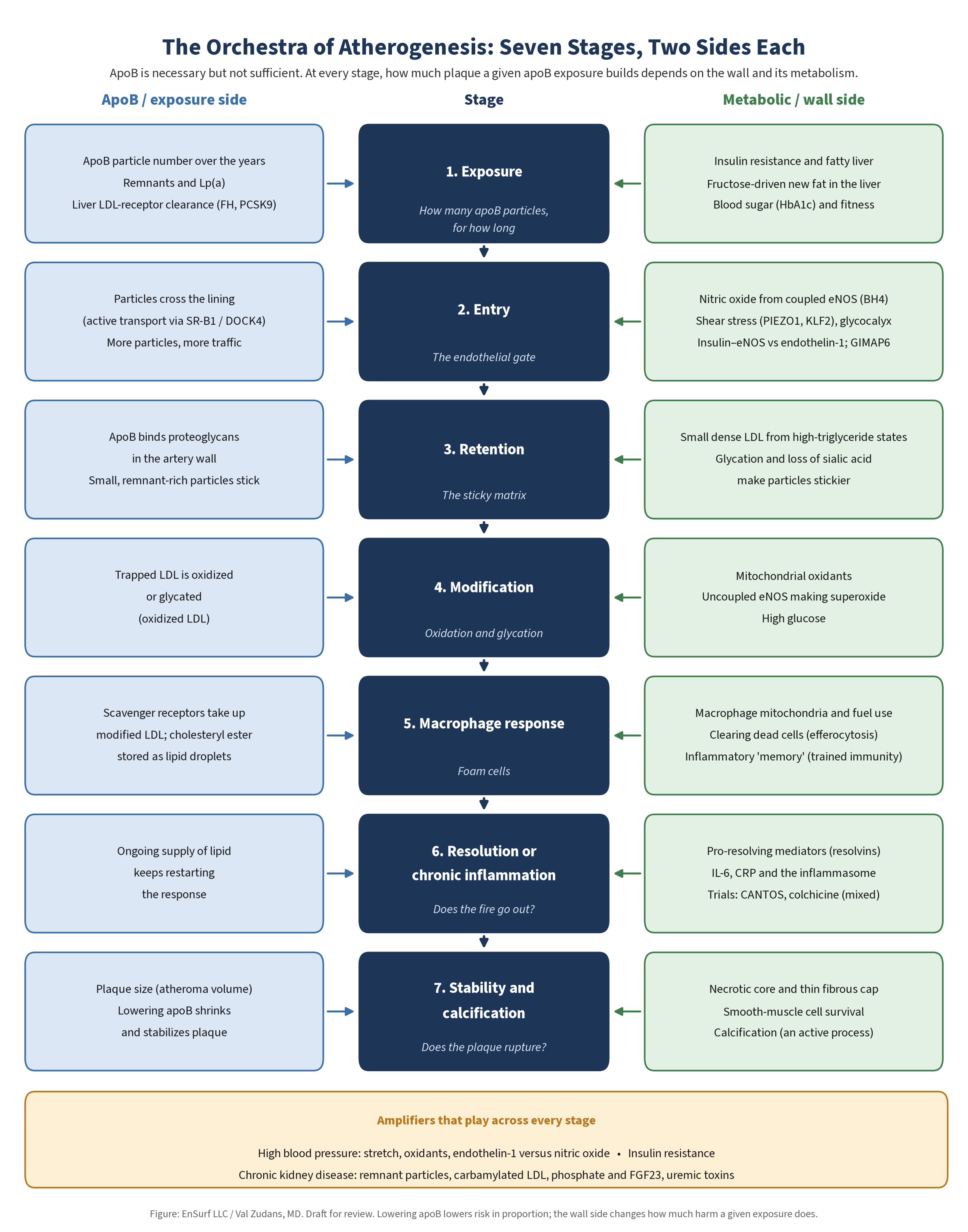
An Orchestra of Atherogenesis: Seven Stages, Two Sides Each
Atherosclerosis is not a solo. It is closer to an orchestra. ApoB particles carry the melody: without apoB-containing lipoproteins there is no lipid core to build, and lowering them lowers risk in proportion [1–3]. But the score is played by many sections at once: the endothelial gate, the matrix beneath it, the redox state of the wall, the macrophages that arrive, the inflammation that either resolves or does not, and the cap that holds the whole thing together. Each of those sections is atherogenic in its own right. Poor metabolic, mitochondrial, and endothelial health is not just background context for apoB. It plays its own part, and it can change how loudly the apoB part is heard.
That is what "necessary but not sufficient" means here. ApoB is necessary: in humans and animals, apoB lipoproteins are the one risk factor that can drive atherosclerosis even when the others are absent [78]. It is not sufficient: how much plaque a given exposure builds, and whether that plaque ever causes an event, depends on the rest of the orchestra. "Necessary" needs a careful reading. The GIMAP6 mice described below developed atherosclerosis with normal, HDL-dominant lipid levels [71]. Their plaques still contained lipid taken up from lipoproteins, so "necessary" here means that some supply of apoB lipoproteins is required, not that apoB must be elevated. How low that requirement goes in a vulnerable wall is not known.
The seven stages below follow a plaque from first exposure to final event. At each one, there is an apoB side and a metabolic or wall side, as Figure 2 shows.
1. Exposure: How Many Particles, for How Long
The apoB side. Exposure is the number of apoB particles in the blood, LDL, remnants, and Lp(a), multiplied by the years they circulate [16, 35, 36]. Lifelong genetic differences in exposure change risk in proportion [1, 3]. Even in middle-aged adults without conventional risk factors, LDL cholesterol in the range usually called normal was independently associated with subclinical plaque [73].
The metabolic side. Metabolic health writes part of the exposure. Insulin resistance drives overproduction of large VLDL, the root of the high-triglyceride, low-HDL, small-dense-LDL pattern [11]. Fructose raises hepatic new-fat production, apoB, and small dense LDL [10]. The liver's cholesterol thermostat sets clearance [41–44]. In the same risk-factor-free adults, glycated hemoglobin was also associated with plaque [73]. The two parts are already playing together before a single particle reaches the wall.
2. Entry: The Endothelial Gate
The apoB side. Particles have to cross the endothelium to start a plaque, and more particles mean more traffic. Entry is not only passive leakage. In mice, LDL is actively carried across endothelial cells by the receptor SR-B1 and its partner DOCK4, and both are increased at lesion-prone sites before plaque forms [79].
The wall side: nitric oxide. The gate is kept by the endothelium, and much of its protection runs through nitric oxide. Endothelial nitric oxide synthase (eNOS) makes NO from L-arginine and oxygen. It needs the cofactor tetrahydrobiopterin (BH4) to do so [80]. NO then diffuses to the smooth muscle beneath, where it activates soluble guanylyl cyclase; the resulting cGMP relaxes the muscle and widens the vessel [80]. NO does far more than dilate. It restrains endothelial adhesion molecules (VCAM-1, E-selectin) and monocyte adhesion [81], reduces platelet adhesion to the endothelium [82], suppresses smooth-muscle growth, and inhibits LDL oxidation [70]. Endothelial dysfunction, with reduced NO activity, is an early marker of atherosclerosis and can be detected before structural changes in the wall are visible [70]. Many of the same risk factors that predispose to atherosclerosis also cause endothelial dysfunction [70].
Figure 3 summarizes the gate.
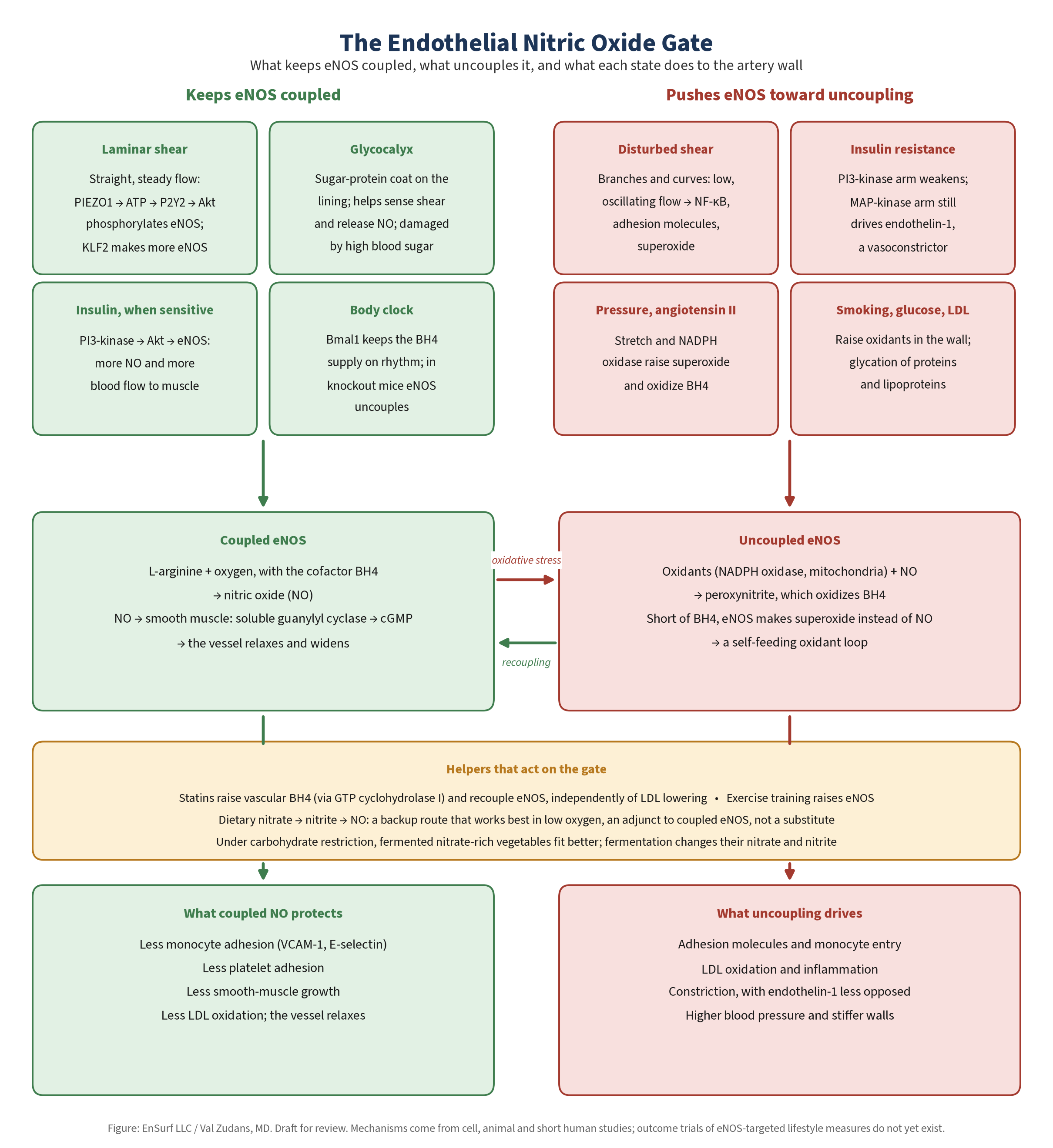
When eNOS fails, it can fail in a particularly harmful way. Hypertension, high cholesterol, diabetes, and smoking raise reactive oxygen species in the vessel wall, largely from NADPH oxidases. Superoxide reacts with NO to form peroxynitrite, which oxidizes BH4. Short of BH4, eNOS becomes "uncoupled" and produces superoxide instead of NO [83]. The enzyme meant to protect the wall becomes a source of the oxidative stress it should prevent. The loop feeds itself, because the superoxide from uncoupled eNOS makes more peroxynitrite, which oxidizes more BH4 [83]. In mice with salt-dependent hypertension, oxidants from NADPH oxidase oxidized BH4 and uncoupled eNOS, and the uncoupled enzyme then became a further source of oxidants [84].
There is a dual face to NO, and it should be stated plainly. High-output inducible NOS, or uncoupled eNOS with abundant superoxide, produces NO and peroxynitrite at levels that injure proteins and mitochondria [83]. On this hypothesis, flooding the system with NO donors is not a clean anti-atherosclerotic strategy. Restoring coupled eNOS, lowering the oxidative load that uncouples it, and keeping shear laminar are the metabolic interventions.
Shear stress. Where blood flows straight and fast, laminar high shear switches on endothelial genes that protect against atherosclerosis. At branches and curves, disturbed, low, oscillating shear switches on genes that promote it, which is why lesions cluster there [85, 86]. Mechanosensing makes this concrete. In mice and endothelial cells, the channel PIEZO1 converts flow into ATP release, purinergic P2Y2 signaling, and phosphorylation of Akt and eNOS; mice lacking endothelial PIEZO1 lost flow-induced NO formation and became hypertensive [87]. Laminar shear also induces the transcription factor KLF2, which raises eNOS and lowers adhesion-molecule expression [88]. Training belongs here: in patients with coronary disease, a few weeks of exercise raised eNOS phosphorylation and improved endothelial function in the internal mammary artery [89].
The glycocalyx. The luminal surface of the endothelium is covered by a mesh of proteoglycans and glycoproteins, the glycocalyx [90]. It contributes to the permeability barrier, inhibits coagulation and leukocyte adhesion, and helps mediate shear-induced NO release [91]. It is proposed to be thinner at lesion-prone branch points [91]. Hyperglycemia damages it: people with type 1 diabetes had reduced glycocalyx volume, worst in those with microalbuminuria [92]. That branch-point thinning helps explain where plaques form is a hypothesis in the reviews cited [91], not a measured human finding.
Hypertension. Pressure and the gate are linked in both directions. Hypertension raises vascular oxidative stress and promotes eNOS uncoupling [83], and loss of endothelial NO signaling can itself raise blood pressure, as in the PIEZO1-deficient mice [87]. The section on hypertension below follows this through every stage.
Insulin resistance and endothelial NO. Insulin has a vascular action as well as a metabolic one. Through the PI3-kinase pathway, it stimulates eNOS and NO-dependent blood flow, which helps deliver glucose to muscle. In insulin resistance, that PI3-kinase branch is selectively impaired while the branch that drives the vasoconstrictor endothelin-1 is not, tipping the endothelium toward constriction [93]. Insulin resistance and endothelial dysfunction then reinforce each other [93].
Clock, fuel, and the nitrate pathway. In mice lacking the core clock gene Bmal1, arteries showed more superoxide, less BH4, and uncoupled eNOS, and adding BH4 improved endothelial function [94]. BH4 levels and the enzymes that make and recycle it followed a daily rhythm in these studies [94], so the clock is part of what keeps eNOS supplied. Whether ordinary circadian disruption in people, from late light, short sleep, or late meals, uncouples eNOS in the same way is not known. Nitrate and nitrite, once seen as inert end products, can be recycled to NO, especially in low-oxygen conditions [95]. Nitrate-rich plants can feed this loop. In keeping with the fermented microbiota-accessible carbohydrates (MACs) of the EnSurf Fuel domain described below, fermented nitrate-rich vegetables fit carbohydrate restriction better than sweet vegetable juices or large servings of raw sweet roots, because lactic acid fermentation uses up part of the sugar in the vegetable [96]. That fit is my dietary judgment, not a tested outcome. Fermentation also changes the nitrogen chemistry, and not in a simple direction. In fermented white and red cabbage and in fermented red beets, nitrate fell during fermentation, and part of it passed into the brine [97]. Nitrite can build up early in a spontaneous ferment, when nitrate-reducing bacteria are active, and fall later as lactic acid bacteria take over [98]. How much nitrate a given fermented vegetable delivers depends on the vegetable, the microbes, and how it is made, and evidence linking fermented beetroot to clinical outcomes is still limited [99]. So fermented vegetables should not be assumed to supply as much nitrate as the same vegetables fresh. On this model, dietary nitrate is an adjunct to coupled eNOS, not a substitute for it: it adds NO without repairing the uncoupled source, and it is a backup route rather than the main one.
Statins at the gate. The interplay shows up even in the drugs. In patients having bypass surgery, a few days of atorvastatin increased vascular BH4, reduced superoxide, and improved NO bioavailability, independently of LDL lowering [100]. In artery segments studied outside the body, atorvastatin raised BH4 by increasing GTP cyclohydrolase I, the enzyme that makes it, which recoupled eNOS; adding back mevalonate reversed the effect, showing that it came from blocking HMG-CoA reductase in the vessel wall itself [100]. An exposure drug also acts on the wall.
Mitochondria and NO. At nanomolar concentrations, NO reversibly inhibits cytochrome oxidase, the last step of the respiratory chain, by competing with oxygen [101]. One proposed role, which is my hypothesis, is that this lets tissue nearest the capillary ration oxygen so it reaches cells farther away. The reversible inhibition itself is measured [101]; whether it works as a tissue-wide matching mechanism in human arteries is not established.
GIMAP6: a wall gene that breaks the gate. A 2026 Cell paper gives the clearest recent example of a wall factor that is atherogenic in its own right [71]. GIMAP6 is an immune-regulatory gene expressed mainly in endothelial cells and macrophages. On a standard C57BL/6 background and an ordinary (standard-fat) diet, mice lacking it developed spontaneous aortic atherosclerosis, coronary disease, myocardial infarction, heart failure, and early death, while keeping a normal, HDL-dominant cholesterol profile with only small changes in triglycerides [71]. A high-fat diet made the plaque worse. Before any plaque was visible, their arteries already leaked, endothelial junctions were disrupted, and monocytes stuck more readily to the endothelium [71].
Crossed onto LDL-receptor-deficient mice, GIMAP6 loss and high LDL acted together, producing advanced, near-occlusive coronary plaque. At comparable plasma cholesterol and triglyceride levels, the double-knockout mice had more atherosclerosis than LDL-receptor-deficient controls on a high-fat diet [71]. In the authors' words, the mechanism "cannot be explained by plasma lipid levels alone."
In humans, rare damaging GIMAP6 variants, pooled across the UK Biobank, All of Us, and Mass General Brigham biobanks, were associated with atherosclerosis but not with high cholesterol, unlike LDLR and PCSK9 variants [71]. A variant enriched in people of African ancestry, V65I, was associated with combined coronary disease and heart failure in All of Us, replicated in the Million Veteran Program, without more hypercholesterolemia. Monocytes from carriers showed less GIMAP6 protein, more CD36, more mitochondrial ROS, and more fatty-acid uptake [71]. All of this rests on a single 2026 study. The mice lacked the gene entirely, on an inbred background, so the work cannot separate what GIMAP6 does in macrophages from what it does in the endothelium. The human associations come from biobank and portal data with limited African-ancestry representation, as the authors note [71].
The lesson for the interplay is not that apoB does not matter. GIMAP6 plaques were built with lipid, and they grew much worse when LDL was high. The lesson is that a failing gate and an overeager macrophage can build plaque at apoB levels that would otherwise be harmless.
3. Retention: The Sticky Matrix
The apoB side. Once inside, apoB particles have to be held. The response-to-retention hypothesis puts this trapping at the center of early disease [102]. ApoB-100 binds negatively charged proteoglycans in the matrix beneath the endothelium. Mice making LDL that could not bind proteoglycans developed much less atherosclerosis, which is direct evidence that retention is an early step [78]. Remnants are retained too, and unlike LDL, their entry into the wall exceeds their exit [9].
The metabolic side. What kind of particle arrives changes how well it sticks. Small dense LDL predicted coronary heart disease in the ARIC study, even in people considered low risk by their LDL cholesterol [103], and small dense LDL is largely a product of insulin resistance and heavy triglyceride traffic [11]. Sugar chemistry matters too. LDL carries sialic acid on its surface, and an enzyme in human plasma can strip it [104]. One research group argues that this desialylation is the earliest and most important step toward the "multiply modified" LDL that loads cells with lipid [105]. This work comes largely from one research group, and its claim that desialylated LDL, rather than oxidized LDL, is the main atherogenic form in human plasma is not established [105].
4. Modification: Oxidation, Glycation, and Redox
The apoB side. Retained LDL does not stay native. Modifications such as oxidation increase its atherogenicity and divert it to macrophage scavenger receptors [106]. Glycated LDL takes a similar path, and small dense LDL is especially prone to glycation [74].
The metabolic side. Modification is a redox event, and the redox state of the wall is a metabolic variable. Excess mitochondrial ROS increases LDL oxidation and endothelial dysfunction, and the classic precursors of atherosclerosis, high cholesterol, high glucose, high triglycerides, and aging, all induce mitochondrial dysfunction [107]. Superoxide from uncoupled eNOS and NADPH oxidases feeds the same pool [83], while coupled NO inhibits LDL oxidation [70].
That does not make antioxidant pills a fix. In a large trial in high-risk patients, vitamin E did not reduce cardiovascular events [108]. Nitric oxide biology draws the same line: the signal is not the enemy; the inefficient, uncoupled source is. Lowering the production of excess oxidants, through coupled mitochondria and coupled eNOS, is a different strategy from mopping them up afterward. That coupling interventions prevent LDL modification in humans is still a hypothesis; the failed antioxidant trials show only that blanket supplementation does not work [108].
5. The Macrophage Response
The apoB side. Retained, modified apoB lipoproteins recruit monocytes. The macrophages they become take up the particles, store cholesteryl ester in lipid droplets, and become foam cells [5, 109]. In a nonresolving response, the lesion grows by cells, lipid, and matrix [109].
The metabolic side. What a macrophage does with that lipid depends on its own metabolism. In the GIMAP6 study, macrophages lacking the gene had higher mitochondrial ROS, which raised PPARγ and the scavenger receptor CD36, lowering the threshold for foam-cell formation even without extra lipid. Blocking mitochondrial ROS with a mitochondria-targeted antioxidant reduced that lipid loading in cells, and in double-knockout mice it reduced plaque and inflammatory cytokines. Deleting CD36 substantially but incompletely rescued early death [71].
Clearing dead cells (efferocytosis) is the macrophage's other job, and it is a mitochondrial one. Macrophages that eat apoptotic cells use the fatty acids from them in mitochondrial β-oxidation and the electron transport chain, with NAD+, to switch on the anti-inflammatory signal IL-10. When their respiratory chain was defective, mice had impaired repair after cardiac injury [110]. In atherosclerosis, efferocytosis is efficient early and fails as plaques advance, and failed efferocytosis is a major driver of the necrotic core [111]. Plaque cells also upregulate CD47, a "don't eat me" signal, and blocking it restored clearance and reduced atherosclerosis in mouse models [112].
Macrophages also remember. Brief exposure of human monocytes to oxidized LDL induced a long-lasting proinflammatory, foam-prone state through epigenetic reprogramming, called trained immunity [113]. In LDL-receptor-deficient mice, a Western diet reprogrammed myeloid progenitors through the NLRP3 inflammasome, and that heightened responsiveness persisted after the mice returned to normal chow [114]. Diet history, not just today's apoB, shapes how the next macrophage behaves. Most of this comes from cell and mouse studies [113, 114]; how much trained immunity adds to human risk is not established.
6. Resolution or Chronic Inflammation
The apoB side. Retained apoB lipoproteins keep feeding the inflammatory response, which is one reason exposure lowering helps at this stage too [109].
The metabolic and immune side. Inflammation is meant to end. The body actively switches it off with specialized pro-resolving mediators, the lipoxins, resolvins, protectins, and maresins, many derived from omega-3 fatty acid pathways [115]. In human carotid plaques, vulnerable regions had lower levels of these mediators relative to the proinflammatory leukotriene LTB4, and giving resolvin D1 to mice promoted more stable plaques with thicker caps and better efferocytosis [116].
Inflammation markers predict events beyond lipids. High-sensitivity CRP was an independent predictor of cardiovascular events in healthy women, and adding inflammatory markers to lipids improved prediction [117]. Human genetics of the IL-6 receptor are consistent with a causal role for IL-6 signaling in coronary disease [72]. Trials now test the lever directly. Canakinumab reduced events without lowering lipids [66]. Low-dose colchicine reduced events after recent myocardial infarction [118] and in chronic coronary disease [119]. But when colchicine was started soon after myocardial infarction in a larger trial, it did not reduce events [120]. So the anti-inflammatory evidence is real but not uniform. The colchicine trials disagree [118–120], and no trial has yet shown that metabolic interventions lower events through inflammation independently of apoB.
7. Stability and Calcification
The apoB side. Most heart attacks come from a plaque that ruptures or erodes, not from slow narrowing, and pathology studies of sudden coronary death have classified the lesions that do this [121]. Lowering apoB can halt or regress atheroma: in pooled serial intravascular ultrasound trials, high-intensity statin therapy was associated with regression of atheroma volume [122].
The wall side. Stability is a property of the wall. Failed efferocytosis enlarges the necrotic core [111], defective resolution thins the cap [116], and mitochondrial dysfunction promotes smooth-muscle apoptosis that weakens it further [107]. Calcification is an active, regulated biological process, not passive mineral deposition [123]. The same statin trials showed coronary calcium increasing even as atheroma regressed [122]. A rising calcium score on a statin is not, by itself, proof that treatment is failing. That reading of statin-associated calcification as stabilization comes from a post hoc analysis of imaging trials [122]; it does not prove that more calcium is protective.
The Score, Read Together
| Stage | ApoB part | Metabolic and wall part |
|---|---|---|
| Exposure | Particle number, remnants, Lp(a), years | Insulin resistance, fructose, liver thermostat |
| Entry | Transcytosis, particle traffic | NO, eNOS coupling, shear, glycocalyx, blood pressure, insulin signaling, GIMAP6 |
| Retention | ApoB-proteoglycan binding | Small dense LDL, glycation, sialylation |
| Modification | Oxidized and glycated LDL | Mitochondrial ROS, uncoupled eNOS |
| Macrophage | Scavenger uptake, cholesteryl ester | Macrophage mitochondria, efferocytosis, trained immunity |
| Resolution | Ongoing lipid feed | Pro-resolving mediators, IL-6, CRP |
| Stability | Atheroma volume | Necrotic core, cap, calcification |
| Blood pressure (every stage) | Stretch speeds particle entry and modification | Oxidants, eNOS uncoupling, endothelin-1 versus NO, the kidney's set point |
| Kidney function (every stage) | Remnant build-up, carbamylated LDL | Phosphate and FGF23, uremic toxins, inflammation, blood pressure |
Read across any row and both parts are playing. The last two rows are amplifiers rather than stages; they are taken up in the two sections that follow. Read down either column alone and you hear only half the score.
Hypertension: Pressure on the Gate
Blood pressure is one of the amplifiers that plays across every stage. In pooled individual data from dozens of prospective studies, the risk of death from heart disease and stroke rose steadily with usual blood pressure throughout middle and old age, with no threshold within the range studied [124]. Lowering pressure lowers events. In a large randomized trial in adults at high cardiovascular risk, a more intensive systolic target reduced a combined outcome of heart attack, stroke, heart failure, and cardiovascular death, and lowered death from any cause, at the cost of more side effects such as low blood pressure, fainting, and kidney injury [125]. Blood-pressure medicines belong with apoB-lowering drugs among the best-proven ways to prevent heart attack and stroke, and nothing here is a reason to stop or change them without your doctor.
How pressure acts on the stages. Hypertension and high cholesterol act on the inner lining of the artery in strikingly similar ways. Both are proinflammatory and recruit monocytes into the intima, probably through oxygen radicals that switch on inflammatory genes [126]. At the entry stage, pressure and stretch raise vascular oxidative stress and push eNOS toward uncoupling [83]. Angiotensin II, a central hormone of blood-pressure control, switches on NADPH oxidase in vascular smooth muscle and raises superoxide [127]. The same oxidants speed the modification of trapped LDL (stage 4), and the inflammatory cells they draw in are the ones that thin the cap of a vulnerable plaque (stages 5 to 7) [126]. In the other direction, losing endothelial NO can itself raise blood pressure, as in mice lacking the endothelial shear sensor PIEZO1 [87].
The nitric oxide and endothelin-1 balance. The endothelium sets vessel tone by balancing two signals. Coupled eNOS makes NO, which relaxes the smooth muscle beneath. Endothelin-1 is a powerful constrictor. Insulin normally drives both, through two branches of its signaling. In insulin resistance, the PI3-kinase branch that drives eNOS weakens while the MAP-kinase branch that drives endothelin-1 does not, tipping the vessel toward constriction, and insulin resistance and endothelial dysfunction then reinforce each other [93]. Salt sits on the same axis. In a small study, salt sensitivity of blood pressure was associated with insulin resistance [128]. In mice on a high-salt diet, vascular superoxide rose and NO fell, and raising the mitochondrial protein UCP2 protected the vessels [129].
The kidney holds the set point. Over the long term, the kidney sets blood pressure through how much salt and water it excretes at a given pressure [130]. That is why the next section, on kidney function, is part of the same story.
My hypothesis of hypertension, in brief. In my metabolic model, sustained high blood pressure is read as the scar of a failed matching system rather than a successful compensation. When the endothelium can no longer match blood flow to tissue energy needs, because eNOS is uncoupled and mitochondria are under strain, the body holds pressure up to keep perfusing underpowered vascular beds, and the kidney, the sympathetic nervous system, and the renin–angiotensin system then lock the higher set point in. That is a hypothesis, not an established mechanism, and it does not change the evidence that lowering blood pressure with proven drugs prevents events [125].
Kidney Function: An Amplifier in Both Directions
Chronic kidney disease is another amplifier that plays across every stage. In a large community-based cohort in an integrated health system, the risks of death and of cardiovascular events rose step by step as estimated kidney function fell, independently of other risk factors [131]. An American Heart Association scientific statement treats kidney disease as a risk factor for the development of cardiovascular disease [132].
Lipoproteins change. Failing kidneys impair the clearance of triglyceride-rich lipoproteins and their atherogenic remnants, so remnant levels rise, and HDL fails to mature, largely because the enzyme LCAT is turned down [133]. On the apoB side, that is more remnant exposure. Uremia also modifies LDL directly. Urea breaks down to cyanate, which carbamylates LDL. In atherosclerosis-prone mice made uremic, carbamylated LDL rose, accumulated in the aortic wall alongside adhesion molecules and macrophages, and went with more severe atherosclerosis [134]. That is a stage-4 modification driven by the kidney, not by apoB number.
Lipid lowering helps, up to a point. In people with moderate to advanced kidney disease, lowering LDL with simvastatin plus ezetimibe reduced major atherosclerotic events [135]. But in two trials in people already on dialysis, starting a statin lowered LDL without significantly reducing the main combined cardiovascular endpoint [136, 137]. The large combined trial found no good evidence that the proportional benefit differed between patients on dialysis and those not on it [135], so the meaning of the dialysis results is debated. One reading, consistent with this essay, is that by the time kidney failure is advanced, much of the risk runs through parts of the orchestra that apoB lowering does not reach. That reading is an interpretation, not a tested result.
Calcification: phosphate and FGF23. Vascular calcification is common in atherosclerosis and a serious problem in kidney disease. In human aortic smooth-muscle cells, phosphate at levels seen in people with high blood phosphate drove mineral deposition and switched on bone-cell genes, through a sodium-dependent phosphate transporter [138]. The body's phosphate hormone, FGF23, rises as the kidneys fail. Higher FGF23 was associated with higher mortality in people starting hemodialysis [139], and in experimental work FGF23 caused pathological growth of heart-muscle cells and was associated with left ventricular hypertrophy in a large kidney-disease cohort [140]. Calcification at stage 7 is therefore partly a mineral-metabolism problem, not only a plaque-healing one.
Inflammation and uremic toxins. In people with advanced kidney failure, carotid atherosclerosis was associated with inflammation, malnutrition, and oxidative stress acting together [141]. Solutes that the failing kidney retains are not inert. Indoxyl sulfate, a protein-bound uremic solute, induces oxidative stress in endothelial cells [142], and indoxyl sulfate, p-cresyl sulfate, and FGF23 have direct toxic effects on the heart [143]. These act at the entry, modification, and resolution stages, on the wall side.
The two-way link with blood pressure. Hypertension and kidney disease feed each other: sustained high pressure worsens kidney function, and declining kidney function worsens blood-pressure control, through fewer nephrons, sodium retention, sympathetic overactivity, the renin–angiotensin–aldosterone system, and endothelial dysfunction [144]. Intensive blood-pressure control reduces cardiovascular events and death in people with kidney disease [144]. On my metabolic model, the kidney's tubules, which spend much of their energy moving sodium, are another high-demand tissue whose mitochondrial strain would show up first as sodium retention and a reset pressure set point. That is a hypothesis.
Kidney disease therefore touches both columns of the score: more remnant and modified particles on the apoB side, and calcification, uremic toxins, inflammation, and pressure on the wall side. Kidney care, blood-pressure care, and lipid care belong together, alongside, not instead of, one another.
Where Each Camp Oversimplifies
Both camps are made up of serious clinicians who have helped patients. The problem is not either camp's core finding. It is the silver bullet each is tempted to extract from it.
The apoB-only simplification: lowering the number is the whole answer. It is a large part of the answer, and for many people, FH above all, the most important part. But treated as the whole answer it neglects insulin resistance, inflammation, fitness, and sleep, which change both vulnerability and apoB itself, and it can leave a patient with a good number and a poor metabolism.
The metabolic-only simplification: apoB doesn't matter if you are metabolically healthy. Metabolic health plausibly lowers vulnerability, and it often lowers apoB. But the genetic evidence says exposure counts in everyone [1, 3], the LMHR data are thin and partly retracted [63–65], and very high diet-induced apoB should not be assumed benign [65]. Treated as a rule, it can lead someone to carry a large exposure for decades on the strength of a hypothesis.
Good management keeps both truths and drops both rules.
Glaucoma: The Same Vulnerability in Another Tissue
Why Glaucoma Belongs in an Essay About Arteries
Glaucoma is a disease of the optic nerve. The retinal ganglion cells that carry vision from the eye to the brain, and their long axons, are lost slowly, often without symptoms until late, and glaucoma is a leading cause of irreversible vision loss worldwide [145]. Lowering eye pressure, with drops, laser, or surgery, is the only treatment proven to slow it [145].
It belongs here for three reasons. First, it shares risk factors with atherosclerosis. Systemic hypertension has a modest association with open-angle glaucoma, and the link is U-shaped: low blood pressure, including low pressure driving blood into the eye and blood-pressure dips at night, is also linked to higher risk, possibly through eye pressure or blood flow to the optic nerve [146]. It is an association, not proven cause. Metabolic syndrome is also associated with a higher risk of glaucoma in pooled observational studies [147]. Second, the eye's drainage tissue runs on the same nitric oxide machinery as the artery. In the conventional outflow pathway, the endothelium lining Schlemm's canal and the smooth-muscle-like cells of the trabecular meshwork use eNOS-derived NO to regulate how easily fluid leaves the eye, and so the eye pressure [148]. Third, statins, the main exposure drug of this essay, have been studied in glaucoma, and their mixed results are a natural test of the difference between exposure and vulnerability.
My Metabolic Hypothesis of Glaucoma, in Brief
I propose that glaucoma and atherosclerosis share an upstream driver: mitochondrial energy failure and redox imbalance in high-demand tissues. This is my hypothesis, and it is not established. Some of its pieces are measured. Trabecular meshwork cells grown from people with primary open-angle glaucoma show a defect in mitochondrial complex I with excess release of reactive oxygen species [149]. Meshwork from these eyes carries more mitochondrial DNA damage than meshwork from control eyes [150]. In a mouse glaucoma model, a subtype of meshwork cells showed mitochondrial dysfunction around the time eye pressure rose, and vitamin B3 (nicotinamide, an NAD+ precursor) protected against the pressure rise [151]. In a small randomized crossover trial in people already on glaucoma treatment, nicotinamide improved a measure of inner retinal function [152]. It is being studied further; it is not a proven treatment and does not replace prescribed drops.
The rest is hypothesis. On this model, the chain described for the artery in the entry stage, oxidative stress, loss of BH4, uncoupled eNOS, and failing NO signaling, would raise outflow resistance in the meshwork, while inflammation and matrix stiffening would lock it in, and retinal ganglion cells, which depend heavily on their mitochondria, would be left with too little energy margin to withstand pressure or poor blood flow. That chain has not been shown in human eyes. Lowering eye pressure stays first, and any change to glaucoma or blood-pressure treatment belongs with your eye doctor and physician.
Statins and Glaucoma: A Test of Exposure Versus Vulnerability
The eye tests the logic. If statins were simply good or bad for energy-hungry neural tissue, the glaucoma data would line up. They do not. A large US managed-care cohort found statin use associated with lower open-angle glaucoma risk [153], and a meta-analysis found a small protective association with short-term use and no clear effect on progression or eye pressure [154]. In the UK Biobank, after adjustment, there was no protective association and no Mendelian-randomization signal [155]. Other cohorts and a network meta-analysis link statins to higher risk. That includes rosuvastatin (hydrophilic) in Australia [156]; rosuvastatin, pravastatin (both hydrophilic), and simvastatin (lipophilic) in a network meta-analysis [157]; and atorvastatin (lipophilic) and rosuvastatin in Taiwan [158]. In the US All of Us cohort, statin use was associated with modestly higher glaucoma prevalence (adjusted odds ratio about 1.13), with stronger associations in some age and LDL-cholesterol strata [159].
So the statin–glaucoma association is inconsistent and is not neatly explained by lipophilicity: hydrophilic and lipophilic statins both turn up on the higher-risk side in some studies. And since CoQ10 depletion is largely systemic [61], tissue penetration is not a clean explanation either. These are observational associations, open to confounding by indication and by who gets eye exams. There is no randomized evidence on statins and glaucoma [154], and none of these associations should change anyone's statin decision on its own.
Two plausible mechanisms pull in opposite directions. In the eye's outflow tissue, statins block the lipid tagging (isoprenylation) that Rho GTPase needs. In pig eyes in organ culture, this relaxed trabecular meshwork cells and increased aqueous outflow, an effect that would lower pressure [160]. That outflow effect comes from experimental organ-culture work. It has not been shown to lower eye pressure in patients, and a meta-analysis found no clear statin effect on eye pressure [154].
In the retina, on the energy-flux hypothesis, systemic CoQ10 depletion could press on retinal ganglion cells, which are high-demand neurons that depend heavily on their mitochondria. That would be a vulnerability effect, not an exposure effect. This is a hypothesis: I found no human study showing that statin-related CoQ10 depletion harms retinal ganglion cell mitochondria.
What the eye does share with the artery is a mitochondrial vulnerability. Trabecular meshwork, retinal ganglion cells, and arterial endothelium are all high-demand tissues with little margin when energy supply falls. In the interplay frame, statins act on exposure; the vulnerability of these tissues is a separate question, and the mixed statin signal says the two should not be collapsed. In glaucoma, as in the artery, proven pressure-lowering and eye-drop therapy stays alongside, not instead of metabolic repair. The same rule applies here to statins and to other LDL-lowering drugs, including FH therapy.
Testing the Interplay
Convergent evidence is consistent with a vulnerability layer. Mitochondrial dysfunction is documented early across vascular cell types. Therapeutic previews—NAD+ precursors, mitochondrial antioxidants, and biogenesis-enhancing exercise—have been proposed to support plaque stabilization and vascular improvement beyond LDL lowering alone, mostly on mechanistic, animal, and small human studies. And reducing inflammation alone reduced events in one large trial [66]. Apart from that anti-inflammatory trial [66], though, the metabolic and mitochondrial levers have no hard-outcome trials showing fewer events independent of apoB. For them, "lowers vulnerability" is a hypothesis, not a result.
The interplay is falsifiable. It predicts that at the same apoB exposure, people with better metabolic and mitochondrial measures will develop less plaque and fewer events, and that metabolic interventions will add benefit on top of apoB lowering. Prospective studies pairing mitochondrial biomarkers (mtDNA heteroplasmy, OXPHOS efficiency) with apoB in LMHR and FH cohorts, or trials of metabolic support plus statin versus statin alone, would test it. If LMHR individuals with sustained high apoB develop plaque at the rate apoB alone predicts, the lower-vulnerability reading fails. If metabolic context makes no difference to outcomes at a given exposure, the vulnerability layer fails, and the apoB-only model is enough. The GIMAP6 findings already point the other way in one monogenic case [71]; the open question is how much common, acquired wall dysfunction, from insulin resistance to uncoupled eNOS, does the same.
Why the Combined Model Explains More
Each camp, alone, explains part of the record. The apoB model explains monogenic FH, the Mendelian randomization results, and the drug trials most directly [1–3, 6]. The metabolic model explains why insulin resistance, hypertension, obesity, and dyslipidemia cluster; why discordant phenotypes exist; why exercise, sleep, and diet move so many markers at once; and why reducing inflammation alone can help [66].
Together they explain more than either does separately: the metabolic syndrome cluster as poor context that also raises exposure; LMHR and carnivore outliers as high exposure in a possibly different context; exercise-induced apoB drops as one lever acting on both; modern dietary harms (chronic insulinogenic overload and, on my hypothesis, seed-oil mitochondrial stress) as evolutionary mismatch acting on context; statin benefits and limits; glaucoma's mixed statin signal as a second high-demand tissue; and parallels with cancer Warburg shifts as primitive compensation for energy deficit. This is not a claim that one camp wins. It is a claim that the full picture needs both.
The lipoprotein life cycle shows the same thing from the inside. Insulin resistance and alcohol raise exposure by loading the liver with fat to export. Low HDL cholesterol is a marker of that context rather than a lever. Lp(a) is exposure that no amount of metabolic repair removes. Each of these sits at a different point on the exposure-vulnerability map, and none is captured by a single number.
Mitochondrial cardiovascular literature is mechanistically rich though historically lighter on large outcomes trials than apoB research. The right posture is to use apoB as a proven target and a valuable signal, and to treat the metabolic layer as the part of the picture that still has to earn its outcome data.
Carbohydrate Restriction: What the Pathways Predict
Carbohydrate restriction acts at the top of the life cycle. Less carbohydrate and less fructose mean lower insulin and less DNL in the liver. In a controlled feeding study, hepatic DNL was much higher on a low-fat, high-carbohydrate diet than on a high-fat, low-carbohydrate one, and it tracked the rise in triglycerides. People with high insulin made more new fat even on the high-fat diet [161]. Less new fat means less triglyceride to export as VLDL. The rest of the cascade follows. Triglycerides fall, HDL cholesterol rises, after-meal lipemia shrinks, and LDL shifts toward larger particles with less small dense LDL [162, 163]. With less triglyceride-rich traffic, there is less CETP exchange to make small dense LDL [11, 17].
At the same time, the liver shifts to burning fat. In obese adults with fatty liver on a short-term, same-calorie, carbohydrate-restricted diet with more protein, liver fat fell quickly, DNL dropped, and ketone (β-hydroxybutyrate) production rose, reflecting more mitochondrial fatty acid oxidation [164]. This is the energy-flux argument in miniature: a liver that oxidizes fat instead of making it.
Insulin resistance is the hub. It links heavy triglyceride traffic, small dense LDL, and low HDL [11] to the vulnerability side: the inflamed, glycation-prone, energy-poor tissue described above. Lowering the insulin load works on both sides of the interplay.
But apoB can move the other way. In lean, healthy young adults, a very-low-carbohydrate, high-fat diet raised LDL cholesterol and apoB on average, with a wide spread between individuals [165]. With moderate carbohydrate restriction, a higher saturated-fat intake offset part of the LDL benefit [163]. This is the hepatic LDL-receptor effect of saturated fat described earlier [46–48], showing up in practice. In lean people on strict carbohydrate restriction, the LMHR pattern of high LDL cholesterol and apoB with low triglycerides and high HDL can appear. The Lipid Energy Model attributes it to higher VLDL export and faster triglyceride uptake in lean, fat-adapted bodies [75, 76]. Both the Lipid Energy Model and LMHR remain hypotheses with limited, partly contested outcome data, and one key LMHR plaque-progression paper has been retracted [63–65]. Nothing here shows that a diet-driven apoB rise is safe.
The practical point is simple. Anyone on a high-saturated-fat, low-carbohydrate diet has good reason to ask their physician about measuring apoB, because some people's rise is large.
Meal timing matters too. In a controlled crossover trial in healthy, normal-weight, middle-aged adults, eating one meal a day without cutting calories raised LDL and HDL cholesterol and blood pressure compared with three meals a day [166]. Compressed eating is not automatically lipid-neutral.
Truth first about FH. Statin-based therapy in FH has decades of outcome evidence and improves survival [6]. Carbohydrate restriction has far less outcome evidence in FH. It may improve context, but it does not fix the underlying LDL-receptor (LDLR) clearance defect, and it is not a substitute for treatment that raises LDL-receptor clearance.
Where This Fits in EnSurf: Tuning the Whole Orchestra, Without Silver Bullets
If exposure and vulnerability interact, management works both levers and fits them to the person. First principles and frugal stewardship point the same way: find out what this person's exposure and context actually are, do the cheap, high-yield work everyone benefits from, and spend on drugs where the evidence says they earn their place. The rest of this section shows how that works in practice.
Lower exposure where it is warranted. Diet can lower apoB. Statins, ezetimibe, PCSK9 inhibitors, and the other LDL-lowering therapies have outcome evidence that the metabolic program does not yet have [2, 6]. They stay alongside metabolic repair, not instead of it, and nothing here is a reason to stop or skip a proven therapy. In FH especially, lifelong lipid lowering is standard care [6]. Knowing that statins work mainly through hepatic LDL receptors [43–45, 49, 50], and that their CoQ10 effect is largely systemic [61], helps a patient and physician choose agent and dose sensibly, not decide whether to treat.
Lower vulnerability. In the EnSurf system, this means three domains, each of which touches the lipoprotein life cycle:
Fuel: Emphasize animal proteins and fats for bioavailable nutrients, include fermented microbiota-accessible carbohydrates (MACs) for microbiome support, use strategic fasting or time-restricted eating, and consider targeted cofactors (PQQ, berberine, NAD precursors, CoQ10, methylene blue in context). These are listed as information only; work with your physician. None has outcome-trial evidence in atherosclerosis, and some interact with prescription medicines. Because saturated fat can turn hepatic LDL receptors down [46–48], anyone who chooses an animal-fat-forward diet should track apoB and decide with their physician what a rise means for them. Lowering refined carbohydrate and fructose works at the top of the cascade, cutting DNL and VLDL triglyceride [10, 161], and the same lever lowers the insulin load that drives vulnerability. Any dose mentioned here is information only, not a prescription.
Low-dose nicotinic acid: information, not a recommendation. Nicotinic acid and nicotinamide are both forms of vitamin B3, but they reach NAD+ by different routes. Nicotinic acid enters the Preiss–Handler pathway through the enzyme NAPRT; nicotinamide is recycled through the salvage pathway, whose key enzyme is NAMPT [167]. Ordinary NAD+ needs can be met from diet alone [168].
At drug doses, nicotinic acid also acts as a drug. It binds the receptor GPR109A (also called HCA2) on fat cells and briefly cuts the release of free fatty acids [169]. That was long thought to explain its lipid effects, but mice lacking the receptor kept the lipid response, and selective receptor agonists lowered free fatty acids in people without niacin-like lipid changes [170, 171]. A more direct liver route is proposed: niacin inhibits DGAT2, an enzyme that makes triglyceride, so more apoB is degraded and fewer VLDL and LDL particles are secreted [172, 173]. At high doses it lowered Lp(a) [174] and raises HDL, partly by slowing the breakdown of apoA-I [173].
Flushing is a separate receptor effect: GPR109A on skin Langerhans cells triggers release of prostaglandin D2 [175, 176]. The threshold varies between people. The US upper intake level for supplemental nicotinic acid is 35 mg a day, a limit a meta-analysis judged conservative [177], and in a small trial, 50 mg twice daily caused flushing in a few patients [178].
Below the flushing range, human lipid data are thin. That trial found a small HDL rise at 50 mg twice daily on top of statins [178], and a dialysis trial that titrated from 25 to 100 mg a day reported a small HDL rise [179]. I found no human lipid study of about 25 mg a day. The idea that a dose this low works mainly as an NAD+ precursor rather than as a lipid drug is my hypothesis, not a tested result.
At gram doses on top of statins, niacin did not reduce events in AIM-HIGH or HPS2-THRIVE, and in HPS2-THRIVE it increased serious problems with diabetes control, muscle, infection, and bleeding [26, 27]. Separately, high blood levels of 2PY and 4PY, end products of excess niacin, were associated with more cardiovascular events, and 4PY raised vascular inflammation in mice; that association generates hypotheses and does not show that niacin intake causes harm [180]. Sustained-release forms caused more liver toxicity than immediate-release niacin [181], and niacin can raise blood glucose modestly [182]. This is information only; any use belongs in a conversation with your physician, especially alongside statins or diabetes medicines.
Circadian: Align light exposure, sleep, and cortisol with evolutionary expectations—morning sunlight, dim evenings, consistent sleep—proposed to support mitochondrial quality control and reduce chronic pseudo-hypoxic signaling. Morning light taken outdoors, in nature, is also the cheapest input on this list. Meal timing belongs here too. In mice, feeding time resets the liver's clock independently of the brain's master clock [183], which is a reason to keep meals regular and daylight-aligned rather than late and scattered.
Strength: Dynamic resistance training is proposed to drive mitochondrial biogenesis and improve insulin sensitivity. In my framework, grip strength and functional capacity serve as practical surrogates of systemic bioenergetic health. Muscle is also where much of the triglyceride in the blood is unloaded. Exercise raises muscle LPL activity [184], and in animal studies inactivity suppresses it quickly [185]. Contracting muscle pulls fuel out of the lipoprotein stream.
Monitoring (information only, with your clinician). ApoB, or non-HDL cholesterol where apoB is not available, tracks exposure [16]. The triglyceride-to-HDL ratio is a simple marker of insulin resistance [186]. Lp(a) is worth measuring once, since it is largely inherited [35]. Fasting insulin, or HOMA-IR calculated from fasting glucose and insulin, gauges insulin resistance [187]. High-sensitivity CRP gauges inflammation, the part of residual risk that CANTOS showed can be treated on its own [66]. Blood pressure and kidney function, already part of routine care, belong on the same list, because both amplify every stage [124, 131]. None of these numbers is a prescription; they are inputs to a conversation with your physician.
Protect the gate. The rule from the entry stage carries over directly: protect the conduit by protecting the source. Anti-atherogenic practice is endothelial-coupling practice. In EnSurf terms, each domain feeds the endothelium as well as the lipoprotein system:
- Fuel. Substrates that mitochondria can oxidize cleanly are proposed to reduce the electron leak and superoxide that uncouple eNOS [83, 107]. Nitrate-rich vegetables can feed the nitrate–nitrite–NO pathway [95], as an adjunct to coupled eNOS, not a substitute for it. Under carbohydrate restriction, fermented nitrate-rich vegetables fit better, since fermentation uses up part of their sugar [96]; fermentation also lowers or shifts their nitrate, so their nitrate content should not be assumed to match fresh vegetables [97, 98]. Lower insulin load protects the PI3-kinase branch of insulin signaling that drives endothelial NO [93].
- Circadian. The vascular clock helps keep eNOS coupled; in mice, losing the clock gene Bmal1 uncoupled it [94]. Regular light, sleep, and meal timing are timing cues for the endothelium as well as the liver.
- Strength and conditioning. Repeated work produces the laminar shear that phosphorylates eNOS today [87, 89] and, through KLF2, writes more of it tomorrow [88]. Training is endothelial gene-expression work as well as mitochondrial biogenesis.
- Resolution. Inflammation that ends is part of the target. Specialized pro-resolving mediators come largely from omega-3 pathways [115], which is one reason fat quality matters beyond apoB. Whether diet or supplements raise these mediators enough to change plaque in humans is not known, so this is a hypothesis, not a protocol.
These Fuel, Circadian, and Strength links to eNOS coupling rest on mechanistic and animal work and on short human studies of endothelial function [83, 87, 89, 93, 94]. None has hard-outcome trial evidence for preventing events.
Do not start with the gain knob. Nitrate donors, PDE5 inhibitors, and even dietary nitrate amplify or supplement NO; they do not replace coupled endothelial production or mitochondrial health. Prescription vasoactive drugs are a physician's decision for specific conditions. Nothing here is a reason to start, stop, or combine them.
Fit the plan to the person. A lean, insulin-sensitive person with a diet-driven apoB rise, an insulin-resistant person with modest apoB and high triglycerides, and a person with FH need different mixes. The first may reasonably weigh a dietary change, the second may gain most from metabolic repair, and the third needs exposure lowering from early in life whatever else they do. In my clinical experience, patients guided this way often achieve meaningful apoB reductions alongside gains in energy, body composition, inflammation, and metabolic flexibility—sometimes with lower medication burden, and sometimes with exactly the drugs they need, used without apology. That is observation from practice, not trial evidence.
Treat the Patient, Not the Lab Value
A lab result measures one person, but the range printed beside it describes a population. Reference intervals are usually set as the central 95% of results from a reference group judged to be healthy [188], and choosing that group, and deciding when age or sex needs its own range, are among the hardest parts of the work [189]. A reference interval is also not the same thing as a clinical decision limit, and confusing the two remains a recognized problem [189]. "Normal" therefore means typical of the reference group. It does not mean optimal, and it does not mean right for this person. This essay has already met that gap: in middle-aged adults without conventional risk factors, LDL cholesterol in the range usually called normal was still associated with subclinical plaque [73].
Risk calculators and treatment targets are models, and models simplify. The Pooled Cohort Equations, used in the United States to estimate a person's risk of a heart attack or stroke, overestimated events in a modern multiethnic cohort; together with three older Framingham-based scores, they overestimated events by 37% to 154% in men and by 8% to 67% in women [190]. In a large, contemporary health-system population of adults without diabetes, the events actually seen were well below those predicted in every risk band, and the miscalibration held across sex, racial and ethnic, and socioeconomic groups; the equation still sorted higher-risk from lower-risk people moderately well, with a C statistic (a measure of that sorting) of 0.74 [191]. Newer equations add kidney function, drop race, and allow optional markers such as urine albumin and HbA1c; in external validation their C statistics were about 0.79 in women and 0.76 in men [192]. That is progress, but any calculator is still built from a handful of inputs. None of them sees the endothelial gate, the mitochondria in the wall, or the rest of the orchestra described here, so a calculator estimate is a starting point for judgment rather than a replacement for it.
Who writes the rules is a fair question too. In a survey of guideline authors, 87% had some tie to the drug industry: 58% had received research funding, 38% had been employees or consultants, and 59% had ties to companies whose drugs the guideline considered. Only 7% thought their own ties had influenced the recommendations, while 19% thought their coauthors' ties had, and specific personal declarations appeared in only 2 of the published guidelines [193]. Among panel members writing lipid and diabetes guidelines in the United States and Canada, 52% had financial conflicts of interest; 11% of those who declared no conflict in fact had one, 6 of the 12 named chairs had conflicts, and conflicts were far less common on government-sponsored panels than on others, 16% versus 69% [194]. In cardiology guidelines, 56% of panel participations involved a reported conflict, ranging from 13% to 87% across guidelines, although many experienced experts on those panels had none [195]. At the level of research, an early systematic review found that industry sponsorship was linked to pro-industry conclusions, with an odds ratio of 3.60 [196]. A later Cochrane review found that industry-sponsored drug and device studies reported favorable efficacy results more often (risk ratio 1.27) and favorable conclusions more often (risk ratio 1.34), and that their conclusions agreed with their own results less often (risk ratio 0.83) than studies with other sponsors [197]. None of this proves that any particular guideline has been captured by industry. It is a reason to ask the question, to expect full disclosure and independent panels, and to read every conclusion against its data.
The opposite trap is just as real. The same Cochrane review found that industry-sponsored studies were not at higher risk of bias on most of the measures it examined, and were more often at low risk of bias from blinding (risk ratio 1.25) [197]. In a randomized study, physicians shown otherwise comparable trial abstracts downgraded a trial's rigor (odds ratio 0.63) and their willingness to prescribe (odds ratio 0.68) simply because industry funding was disclosed, and were about half as willing to prescribe a drug tested in an industry-funded trial as one tested in an NIH-funded trial (odds ratio 0.52), even when the trial was well designed [198]. The PCSK9-inhibitor outcome trial cited in this essay was funded by its manufacturer [2], and its result agrees with genetic evidence that lower LDL exposure lowers risk [1, 3]. Judge a study first by its design and its data, then weigh who paid for it; do not reject good evidence because of the sponsor alone.
In practice. The same apoB number can mean different things in different people. A level that is acceptable for one person may be too high for someone with FH, a high Lp(a), kidney disease, diabetes, or a family history of early heart disease, and a coronary artery calcium scan, where appropriate, can show whether exposure has already built plaque. The current cholesterol guideline itself names such risk-enhancing factors, uses coronary artery calcium scoring to help when a treatment decision is uncertain, and calls for a clinician–patient risk discussion and shared decision-making [199]. Blood pressure and kidney function amplify every stage [124, 131], and metabolic markers such as triglycerides, HbA1c, and measures of insulin resistance help show how vulnerable the wall is likely to be. Read together with a clinician, the whole picture tells a person where they stand: how much to lower exposure, and how much work the wall and metabolism need. That is the thesis of this essay applied to one patient: lower exposure where it is warranted, lower vulnerability, and fit both to the person.
None of this is advice to ignore lab results or to stop medicines. A result outside the range, or a calculator estimate that seems wrong for you, is something to discuss with your clinician, not a reason to change treatment on your own.
Conclusion
Follow one apoB particle from the liver to the artery wall and both camps turn out to be describing the same system. The liver assembles VLDL from fat supplied by the diet, by adipose tissue, and by new fat made from carbohydrate. LPL in muscle and fat unloads the triglyceride, hepatic lipase and CETP remodel what is left, and the liver's LDL receptors decide how long the remnants and LDL stay in the blood. HDL runs a separate track that pulls cholesterol back out of cells. Alcohol, fructose, and insulin resistance load the liver with fat to export; statins, through SREBP2, make the liver clear more particles; saturated fat can turn those receptors down; Lp(a) is set at birth.
Exposure, the number of apoB particles and how long they circulate, is necessary but not sufficient. Vulnerability, the metabolic and mitochondrial state of the wall that receives them, decides how much damage a given exposure does, and the same metabolic state helps set the exposure in the first place. Insulin resistance is the hub where the two meet. The eye's mixed statin signal is a reminder that exposure and vulnerability are different questions, even when one drug touches both.
The stages of plaque-building make the same point in more detail. At every step, from exposure and entry through retention, modification, the macrophage response, resolution, and stability, there is an apoB part and a metabolic or wall part. Nitric oxide and the endothelial gate, the glycocalyx and shear, insulin signaling, mitochondrial ROS, efferocytosis, trained immunity, and pro-resolving mediators are not footnotes to apoB. They are sections of the same orchestra, and GIMAP6 shows that a single wall gene can make that orchestra play loudly with normal cholesterol. High blood pressure and failing kidneys amplify every stage, on both sides of the score. ApoB is necessary but not sufficient. Lowering it lowers risk in proportion, and that evidence stands. Tuning the rest of the orchestra is the other half of the job.
So the practical lesson is to stop looking for silver bullets. Lowering the number is not the whole answer, and metabolic health is not a licence to ignore the number. In the EnSurf frame, Fuel, Circadian, and Strength lower vulnerability and often lower exposure too; measuring apoB, Lp(a), and insulin resistance shows where a given person actually stands; and statins and other LDL-lowering therapies, including FH therapy, stay alongside, not instead of, that work when exposure needs lowering. Fit the mix to the person in front of you. That is the frugal, first-principles path, and it is more likely to narrow the residual-risk gap than either camp's rule alone.
This is still a hypothesis. It earns its keep only if the tests above come out the way it predicts.
This essay is a hypothesis. It draws from peer-reviewed mechanisms in mitochondrial biology and vascular pathology, phenotypic observations, and clinical patterns as of 2026. It is not medical advice and is not a reason to stop any prescribed therapy. Individual care requires personalized evaluation by a qualified physician.
2b. Nitric Oxide — energy flux manager
Hypothesis language only — not a nitrate/PDE5 protocol, not a blood-pressure drug holiday, not an erectile self-treatment guide. Do not start, stop, or combine nitrates, PDE5 inhibitors, or other vasoactive drugs from this page. Education only; no Rx advice.
Nitric oxide is conventionally taught as a vasodilator. That description is true and incomplete. NO is better understood as an energy-flux manager: a short-lived gas that simultaneously adjusts delivery of oxygen and substrate to working mitochondria, rations consumption of oxygen at cytochrome c oxidase so proximal organelles do not steal the entire gradient, and protects the conduit so that future flux is not obstructed by adhesion, thrombosis, and smooth-muscle overgrowth. When NO bioavailability is adequate and coupled, high energy flux is possible without proportional damage. When it is lost, the cell experiences a delivery-and-matching failure that the mitochondrial-metabolic hypotheses of aging and cancer already identify as upstream: chronic mismatch between ATP supply and maintenance demand.
Full essay: Sage — The Metabolic Hypothesis of Nitric Oxide.
Glaucoma: Three Compartments, One Matching Failure
Glaucoma has its own page: Sage — Glaucoma. That page covers the three compartments and what nitric oxide does in glaucoma.
PDE5 Inhibitors: Amplifier, Not Source
The vascular pharmacology of the class: Sage — Nitric Oxide.
The nitric oxide profile described above has two ocular chapters that must not be collapsed into one.
Cyanopsia---the blue color shift---is PDE6. Photoreceptor PDE6 hydrolyzes cGMP after a photon hits the opsin. That drop closes cation channels and starts the visual signal. Sildenafil is only about ten-fold selective for PDE5 over PDE6; tadalafil is far more selective. Partial PDE6 block leaves cGMP a little high after a flash, slows phototransduction, and impairs blue--green discrimination. Patients report a blue tinge, extra brightness, photophobia. It tracks plasma peak and clears in hours. It is not optic-nerve ischemia, and it is not known to predict NAION.
NAION is a perfusion-compartment event in a crowded disc. The optic nerve head sits in a tight scleral canal, supplied by short posterior ciliary arteries. Most patients who infarct that tissue already have a small cup-to-disc ratio---"disc at risk." A transient drop in perfusion pressure (mean arterial pressure minus intraocular pressure) below what that anatomy can tolerate produces axoplasmic stasis, swelling, and further compression. Many ordinary NAION cases are noticed on waking, and nocturnal hypotension has been proposed as a final insult.
A PDE5 inhibitor can be the last increment: mild systemic vasodilation, a nocturnal pressure dip after an evening dose, perhaps a small transient IOP rise. The drug does not clog the nerve. It lowers the pressure head in a nerve that had almost no anatomic reserve. Labels advise caution in anyone who has already had NAION, because recurrence risk is higher, and tell anyone with sudden vision loss to stop the drug and seek care. Absolute risk is low. Relative risk in selected observational analyses is on the order of twofold on a background of about 1 case per 10,000 person-years in people over 50. Causality is plausible, not a large population effect.
Keep the chapters separate. Blue vision is homology between two cGMP phosphodiesterases. NAION is a rare ischemic event in a structurally vulnerable nerve plus a blood-pressure dip. Neither is an argument for treating every optimized adult with daily tadalafil. Both are arguments for knowing the anatomy you are asking a drug to visit. The same anatomy---crowded disc, nocturnal dip, perfusion pressure as MAP minus IOP---is the acute version of the matching failure that, left chronic, is one corridor into glaucomatous nerve-head damage.
Vascular/cardiac depth lives on this page. Signaling-nest framing (NO beside adenosine / insulin as matching loops) also appears on Sage — Signals. Ocular shear–eNOS / glaucoma compartment language cross-links Sage — Eye.
3. Autonomic / POTS — orthostatic expression of constrained energy economy
POTS is conventionally framed as a primary disorder of autonomic regulation. Diagnostic criteria center on an excessive heart-rate rise upon standing (≥30 bpm adults / ≥40 bpm adolescents) without orthostatic hypotension, with chronic orthostatic intolerance that improves on recumbency. Subtypes (hypovolemic, neuropathic, hyperadrenergic, deconditioning-dominant) and algorithms emphasizing volume, compression, graded exercise, and symptom-targeted pharmacology are clinically useful — and incomplete. On the metabolic hypothesis, they describe a compensatory surface while leaving the possible energetic substrate incompletely examined. [POTS 1]
The metabolic hypothesis reframes POTS as the orthostatic expression of constrained cellular energy economy. In this model, tachycardia can be compensatory: the system’s attempt to preserve cerebral perfusion and cardiac output when stroke volume falls and energy-dependent vascular and neural control falter under gravitational load. The hypothesis proposes that mitochondrial inefficiency — potentially unmasked or induced by infection — could reduce reserve in the tissues that matter most under orthostasis: vascular smooth muscle, autonomic nerves, cardiac myocytes, and the brain. Relative hypovolemia, impaired vasoconstriction, exaggerated sympathetic drive, and cardiac atrophy could, in this model, emerge as interlocking downstream consequences rather than necessarily independent primary lesions.
Upon standing, hundreds of milliliters of blood pool in the lower body and splanchnic bed. Venous return declines, stroke volume drops, and baroreceptor unloading triggers compensatory tachycardia and vasoconstriction. In a metabolically robust individual the response is modest and effective. In the POTS patient the same gravitational stress elicits an exaggerated rate response because the capacity to maintain stroke volume and peripheral resistance has been compromised; an energetic contribution is proposed here. [POTS 1] [POTS 2] High-demand tissues cannot maintain membrane potential, calcium handling, or contractile tone when oxidative phosphorylation is impaired. Cerebral neurons are exquisitely sensitive to modest energy shortfalls — brain fog and near-syncope follow.
Preliminary evidence cluster from different clinical and experimental settings (hypothesis-relevant, not confirmation or a protocol): fibroblast findings linking choline/betaine handling, membrane phospholipid ratios, oxygen consumption, membrane potential, and ROS; primary mitochondrial cytopathies producing orthostatic intolerance; overlap with ME/CFS energetics; post-viral (including long COVID) mitochondrial disruption — Complex I vulnerability, ROS cycles, NAD⁺ drain, fragile mitophagy, altered inner-membrane phospholipids. In this hypothesis, subtypes may be overlapping expressions of an energetic constraint: relative hypovolemia because volume regulation costs energy; neuropathic features as small-fiber/ganglia energy failure; hyperadrenergic drive as a potentially compensatory sympathetic response to inadequate output or cellular supply; deconditioning as both consequence and amplifier. Autoimmunity and unresolved post-viral immune activity are interpreted here as potentially energetically expensive compensations when ATP capacity is short of allostatic load. [POTS 3] [POTS 4] [POTS 5] [POTS 6] [POTS 7] [POTS 8]
Therapeutic logic in the Sage essay follows hierarchy without converting this Sage into orders: expand plasma volume and reduce pooling; progressive initially-recumbent reconditioning; substrate selection that favors metabolic flexibility and lowers peroxidation load; circadian alignment as non-negotiable autonomic stabilizer; wearable HRV/resting HR and grip as recovery dashboards; pharmacologic agents as temporary scaffolding while capacity rebuilds. Targeted mitochondrial cofactors appear in the essays as rate-limiting supports applied to documented deficits — still hypothesis language under medical supervision, not a retail dosing protocol. This page does not invent peptide, HRT, or GLP-1 regimens. [POTS 10] [POTS 11]
POTS is heterogeneous. The hypothesis does not require uniformity; it requires that cellular energy economy is a rate-limiting node in the majority and that addressing it can produce meaningful gains. In cascade vocabulary: orthostatic tachycardia is acute-looking compensation that becomes a chronic emergency phenotype when reserve never returns.
4. Organ dysfunction — multi-organ cardiac angle
From The Metabolic Hypothesis of Organ Dysfunction: organs are integrated tissue assemblies where specialized cells perform high-work functions supported by vascular, matrix, and local signaling networks. When level 0–1 ATP / redox homeostasis fails in key cell populations — fuel inefficiency (including peroxidized cardiolipin, proposed here to come from seed-oil PUFAs), hyperglycemia, cofactor gaps, circadian misalignment (which in mice disrupts PGC-1α-linked rhythms), ROS, mtDNA damage, impaired mitophagy — higher-level responses may activate: vascular remodeling, matrix reorganization, altered fluid dynamics, local immune clearance, functional adaptation or sacrifice of vulnerable cell types. In this model, acute activation can be adaptive; chronic persistence would reflect failed resolution.
The heart is named explicitly as a parallel organ of force equal to the ocular archetype. Cardiomyocytes possess a very high mitochondrial volume density to support continuous contractile work. Chronic level 0–1 insufficiency — from hypertension-induced pressure overload, ischemic episodes, diabetic glucolipotoxicity, or dietary insults that impair mitochondrial handling — may trigger compensatory hypertrophy to normalize wall stress despite inefficient sarcomere force generation; extracellular matrix remodeling and fibrosis may represent repair attempts that eventually stiffen the ventricle (HFpEF patterns) or dilate it (HFrEF patterns). Benefits of agents such as SGLT2 inhibitors in heart failure are read, in Organ Dysfunction framing, as possibly including favorable shifts in myocardial fuel utilization and reduced glucotoxicity on mitochondria — consistent with upstream support — without converting this essay into a prescribing guide.
Kidney and brain parallels matter for cardiology multimorbidity: proximal tubular / podocyte energy failure contributing to impaired reabsorption, proteinuria, scarring compensations; neuronal bioenergetic decline → protein mishandling, synaptic loss, gliosis, vascular cognitive impairment. Inflammaging and microglial activation as chronic clearance attempts mirror vessel-wall inflammatory persistence. Atherosclerosis and myocardial energy failure are therefore not separate ontologies. Vessel-wall bioenergetic collapse and myocyte bioenergetic collapse are tissue expressions of the same mismatch geometry under different clocks and loads. The eye remains the clinical window; the heart is the continuous engine under the same rule. Specialty cardiology owns minutes, ischemia, arrhythmia salvage, and procedure clocks. This Sage owns question order: engine before dashboard theater.
Cardiomyopathy and heart failure in depth: Sage — Cardiomyopathy and Heart Failure.
5. Hypertension — scar of matching failure
Hypertension-as-scar is covered in Sage — Nitric Oxide. That essay supplies the EnSurf-compatible account: chronic hypertension is not successful ATP compensation via higher cuff pressure; it is cross-system scar — uncoupled eNOS, high standing resistance, wall remodeling, and renal pressure-natriuresis reset — after matching loops were left on as architecture. This does not invent a BP-drug holiday or a Surfer antihypertensive protocol. Specialty care still owns pressure emergencies and indicated pharmacotherapy.
Primary hypertension in depth: Sage — A Metabolic Hypothesis of Hypertension.
Earlier peripheral mentions remain true and subordinate: The Good Doctor lists essential hypertension among chronic clinic faces of compensations that did not stand down; Organ Dysfunction treats hypertension-induced pressure overload as one driver of myocardial level 0–1 insufficiency.
6. Cardiac electrophysiology
Electrophysiology and arrhythmia in depth: Sage — Electrophysiology and Arrhythmia.
Bridge logic:
- Ion pumps are ATP machines. Resting membrane potential, Na⁺/K⁺-ATPase work, and sarcoplasmic reticulum calcium cycling are among the most expensive continuous costs in excitable tissue. When mitochondrial ATP supply and redox order falter, excitability and conduction become less stable — the same geometry POTS already states for membrane potential and calcium handling in myocytes, smooth muscle, and autonomic axons under orthostatic load.
- Adenosine as energy-stress dashboard. The Adenosine Energy Stress Signal essay frames adenosine/AMP as the high-frequency signal of ATP breakdown — protective braking and demand reduction when supply is short. That is dashboard language for energy stress in excitable systems, not an antiarrhythmic protocol and not a claim that every clinical arrhythmia is “only adenosine.”
- Chronic emergency-mode. On the compensatory cascade, acute lower-nest compensation can preserve persistence; chronic lock-in (glycolytic bias, ROS, membrane lipid injury from Fuel mismatch) is the hypothesized substrate in which ectopy, conduction delay, or autonomic rate extremes become more likely. Plaque ischemia and myocyte energy failure (Organ Dysfunction) remain classic structural routes into the same phenotype space.
What this section refuses: fabricated EP trial citations; invented ablation/drug algorithms; peptide or hormone “rhythm protocols”; any implication that Fuel/light/strength coaching replaces ICD, anticoagulation, or EP lab timing. Specialty electrophysiology owns those clocks. It asks the Good Doctor’s question in a new tissue: is ATP-generating capacity sufficient and flexible for the electrical work this heart is being asked to do?
7. Conventional alternatives / Surf Judge — Good Doctor
Conventional cardiology has produced remarkable saves: reperfusion, devices, antiplatelet and anticoagulant pathways, pressure and rate control, lipid-lowering where indicated, structured rehab, HF drug classes that improve outcomes. The metabolic map does not compete with those minutes. It relocates question order.
Map ≠ terrain. LDL/ApoB, Lp(a), blood pressure, heart-rate rise on stand, EF, calcium score, wearable HRV — these are indispensable dashboards. They are not the organism’s Level 0–1 field. Treating the dashboard as the disease while the mismatch continues is the Good Doctor’s named error mode. Treating the constraint (restore oxidative capacity and fuel-handling resilience; reduce chronic emergency demand) is complementarity, not ideology against stents.
Therapeutic implication language already in the Lp(a) addendum and Organ Dysfunction: emerging Lp(a)-specific lowering can reduce OxPL-laden cargo influx where specialty care judges it indicated; mitochondrial support and board work address handling capacity; SGLT2i benefits in HF are readable partly as fuel-utilization shifts. EnSurf pillar translation remains hypothesis, not cardiac rehab orders:
- Fuel: nutrient-dense patterns supporting beta-oxidation and TCA flux; avoid chronic hyperinsulinemic and seed-oil–dominant patterns that impair mitochondrial membrane handling — same Fuel geometry as other Sage pages.
- Circadian: morning outdoor light, consistent sleep, dim evenings — support mitochondrial quality-control and autonomic stability; chaotic nights are engine noise.
- Strength: progressive resistance and cardiorespiratory capacity as biogenesis stimuli; grip and fitness as honest systemic dashboards; for POTS, the essay emphasizes progressive, initially recumbent loading.
None of this authorizes stopping cardiology meds from a webpage, delaying stents or thrombolysis, coaching through syncope as a fasting experiment.
8. Cross-links
- Fuel — membrane peroxidation, substrate architecture, vascular injury pathway.
- Nitric Oxide (this page §2b) — 2.0 flux manager / scar grammar; Education only; no Rx advice.
- Signals — hormones/peptides as higher coordination; NO matching-loop nest beside adenosine/insulin (no invented Rx / PDE5 protocols).
- Serotonin — sibling flux manager (oxygen/carbon allocation); no Coach invention.
- Thyroid — non-thyroidal illness / conservation dashboard under energy constraint; shared cascade geometry.
- Aging — progressive ATP-capacity loss as shared spine.
- Immune / inflammation — inflammatory persistence as compensation that never stood down; athero convergence.
- Diabetes · Obesity — glucolipotoxicity and diabesity field for vessel and myocardium.
- Eye — organ-dysfunction archetype; heart named as equal-force parallel.
- Philosophy of Medicine — disease as chronic compensatory mode; Good Doctor ethos.
- Methylene Blue — electron-carrier hypothesis; appears in the POTS essay as one researched support under supervision — not a cardiology cure claim here.
9. Limits of the claim
- Working hypothesis. Not a randomized-trial verdict that mitochondrial-first strategies outperform guideline ApoB lowering, HF guidelines, or autonomic specialty algorithms as monotherapy.
- Does not deny ApoB biology, OxPL, thrombosis, ischemia, structural heart disease, or the clinical utility of lipid-lowering, antiplatelet, antihypertensive, rate/rhythm, revascularization, or device care when indicated.
- FH, elevated Lp(a), established ASCVD, syncope, and acute coronary syndromes remain high-stakes; specialty care owns those clocks.
- Not covered here: valve disease. Ocular NO detail lives on Eye Sage.
10. Conclusion
Cardiology, in the Metabolic Hypothesis map, is what chronic emergency-mode metabolism looks like when the mismatch is written into vessel wall, myocardium, autonomic orthostatic control — and, as a proposed bridge, into the ATP-expensive electrical work of excitability and conduction. Atherosclerosis is more fundamentally a disorder of cellular energy handling in the vascular wall than of apoB retention alone — apoB particles, oxidized cargo, and Lp(a) as amplifiers of an upstream mitochondrial-metabolic crisis. On this hypothesis, POTS may include orthostatic compensation when mitochondrial capacity is insufficient for upright posture. Heart failure patterns are organ-level hypertrophy/fibrosis records of myocyte energy failure. Hypertension is now mapped as NO-matching scar, not as successful pressure compensation. Electrophysiology is invited into the same question order. Lipids, rate, pressure, and wearable scores are dashboards; mitochondria and fuel architecture are engine. The Good Doctor begins with persistence: whether ATP-generating capacity is sufficient and flexible for demand. Coach and Surfer give shorter versions. Cardiology still owns the emergency.
Topics in this chapter
- Atherosclerosis
- Nitric Oxide — NO flux manager / scar grammar
- Hypertension — pressure as readout, resistance as scar (hypothesis)
- Autonomic / POTS — orthostatic expression of constrained energy economy
- Organ dysfunction — multi-organ cardiac angle
- Cardiomyopathy and Heart Failure — chronic energy starvation under sustained load (hypothesis)
- Cardiac electrophysiology
- Electrophysiology and Arrhythmia — energy-dependent ion homeostasis (hypothesis)
- Glaucoma
- Pulmonary — the oxidizing-magnet half of the dual-delivery architecture (hypothesis)
- Arterial and Venous Occlusions — retinal, coronary, cerebral, and venous occlusion as one upstream failure (hypothesis; home: Ophthalmology)
A working hypothesis, open to correction and revised as new evidence arrives. Last revised November 6, 2026.
References
2. Atherosclerosis — ApoB and the Engine: An Orchestra of Atherogenesis
- Ference BA, Ginsberg HN, Graham I, et al. Low-density lipoproteins cause atherosclerotic cardiovascular disease. 1. Evidence from genetic, epidemiologic, and clinical studies. A consensus statement from the European Atherosclerosis Society Consensus Panel. Eur Heart J. 2017;38:2459–2472. doi:10.1093/eurheartj/ehx144. PMID 28444290.
- Sabatine MS, Giugliano RP, Keech AC, et al. Evolocumab and clinical outcomes in patients with cardiovascular disease (FOURIER). N Engl J Med. 2017;376:1713–1722. doi:10.1056/NEJMoa1615664. PMID 28304224.
- Ference BA, Kastelein JJP, Ray KK, et al. Association of triglyceride-lowering LPL variants and LDL-C-lowering LDLR variants with risk of coronary heart disease. JAMA. 2019;321:364–373. doi:10.1001/jama.2018.20045. PMID 30694319.
- Brown MS, Goldstein JL. Lipoprotein metabolism in the macrophage: implications for cholesterol deposition in atherosclerosis. Annu Rev Biochem. 1983;52:223–261. doi:10.1146/annurev.bi.52.070183.001255. PMID 6311077.
- Ghosh S, Zhao B, Bie J, Song J. Macrophage cholesteryl ester mobilization and atherosclerosis. Vascul Pharmacol. 2010;52:1–10. doi:10.1016/j.vph.2009.10.002. PMID 19878739.
- Defesche JC, Gidding SS, Harada-Shiba M, et al. Familial hypercholesterolaemia. Nat Rev Dis Primers. 2017;3:17093. doi:10.1038/nrdp.2017.93. PMID 29219151.
- Hussain MM, Rava P, Walsh M, et al. Multiple functions of microsomal triglyceride transfer protein. Nutr Metab (Lond). 2012;9:14. doi:10.1186/1743-7075-9-14. PMID 22353470.
- Donnelly KL, Smith CI, Schwarzenberg SJ, et al. Sources of fatty acids stored in liver and secreted via lipoproteins in patients with nonalcoholic fatty liver disease. J Clin Invest. 2005;115:1343-51. doi:10.1172/JCI23621. PMID 15864352.
- Ginsberg HN, Packard CJ, Chapman MJ, et al. Triglyceride-rich lipoproteins and their remnants: metabolic insights, role in atherosclerotic cardiovascular disease, and emerging therapeutic strategies-a consensus statement from the European Atherosclerosis Society. Eur Heart J. 2021;42:4791-4806. doi:10.1093/eurheartj/ehab551. PMID 34472586.
- Stanhope KL, Schwarz JM, Keim NL, et al. Consuming fructose-sweetened, not glucose-sweetened, beverages increases visceral adiposity and lipids and decreases insulin sensitivity in overweight/obese humans. J Clin Invest. 2009;119:1322-34. doi:10.1172/JCI37385. PMID 19381015.
- Adiels M, Olofsson SO, Taskinen MR, et al. Overproduction of very low-density lipoproteins is the hallmark of the dyslipidemia in the metabolic syndrome. Arterioscler Thromb Vasc Biol. 2008;28:1225-36. doi:10.1161/ATVBAHA.107.160192. PMID 18565848.
- Kersten S. Physiological regulation of lipoprotein lipase. Biochim Biophys Acta. 2014;1841:919-33. doi:10.1016/j.bbalip.2014.03.013. PMID 24721265.
- Zhang R. The ANGPTL3-4-8 model, a molecular mechanism for triglyceride trafficking. Open Biol. 2016;6:150272. doi:10.1098/rsob.150272. PMID 27053679.
- Mahley RW, Huang Y. Atherogenic remnant lipoproteins: role for proteoglycans in trapping, transferring, and internalizing. J Clin Invest. 2007;117:94-8. doi:10.1172/JCI30889. PMID 17200713.
- Santamarina-Fojo S, González-Navarro H, Freeman L, et al. Hepatic lipase, lipoprotein metabolism, and atherogenesis. Arterioscler Thromb Vasc Biol. 2004;24:1750-4. doi:10.1161/01.ATV.0000140818.00570.2d. PMID 15284087.
- Sniderman AD, Thanassoulis G, Glavinovic T, et al. Apolipoprotein B Particles and Cardiovascular Disease: A Narrative Review. JAMA Cardiol. 2019;4:1287-1295. doi:10.1001/jamacardio.2019.3780. PMID 31642874.
- Barter PJ, Brewer HB Jr, Chapman MJ, et al. Cholesteryl ester transfer protein: a novel target for raising HDL and inhibiting atherosclerosis. Arterioscler Thromb Vasc Biol. 2003;23:160-7. doi:10.1161/01.atv.0000054658.91146.64. PMID 12588754.
- Jiang XC, Yu Y. The Role of Phospholipid Transfer Protein in the Development of Atherosclerosis. Curr Atheroscler Rep. 2021;23:9. doi:10.1007/s11883-021-00907-6. PMID 33496859.
- Barter PJ, Caulfield M, Eriksson M, et al. Effects of torcetrapib in patients at high risk for coronary events. N Engl J Med. 2007;357:2109-22. doi:10.1056/NEJMoa0706628. PMID 17984165. (ILLUMINATE)
- Lincoff AM, Nicholls SJ, Riesmeyer JS, et al. Evacetrapib and Cardiovascular Outcomes in High-Risk Vascular Disease. N Engl J Med. 2017;376:1933-1942. doi:10.1056/NEJMoa1609581. PMID 28514624. (ACCELERATE)
- Bowman L, Hopewell JC, Chen F, et al. Effects of Anacetrapib in Patients with Atherosclerotic Vascular Disease. N Engl J Med. 2017;377:1217-1227. doi:10.1056/NEJMoa1706444. PMID 28847206. (HPS3/TIMI55–REVEAL)
- Rosenson RS, Brewer HB Jr, Davidson WS, et al. Cholesterol efflux and atheroprotection: advancing the concept of reverse cholesterol transport. Circulation. 2012;125:1905-19. doi:10.1161/CIRCULATIONAHA.111.066589. PMID 22508840.
- Yvan-Charvet L, Ranalletta M, Wang N, et al. Combined deficiency of ABCA1 and ABCG1 promotes foam cell accumulation and accelerates atherosclerosis in mice. J Clin Invest. 2007;117:3900-8. doi:10.1172/JCI33372. PMID 17992262.
- Khera AV, Cuchel M, de la Llera-Moya M, et al. Cholesterol efflux capacity, high-density lipoprotein function, and atherosclerosis. N Engl J Med. 2011;364:127-35. doi:10.1056/NEJMoa1001689. PMID 21226578.
- Voight BF, Peloso GM, Orho-Melander M, et al. Plasma HDL cholesterol and risk of myocardial infarction: a mendelian randomisation study. Lancet. 2012;380:572-80. doi:10.1016/S0140-6736(12)60312-2. PMID 22607825.
- Boden WE, Probstfield JL, Anderson T, et al. Niacin in patients with low HDL cholesterol levels receiving intensive statin therapy. N Engl J Med. 2011;365:2255-67. doi:10.1056/NEJMoa1107579. PMID 22085343. (AIM-HIGH)
- Landray MJ, Haynes R, Hopewell JC, et al. Effects of extended-release niacin with laropiprant in high-risk patients. N Engl J Med. 2014;371:203-12. doi:10.1056/NEJMoa1300955. PMID 25014686. (HPS2-THRIVE)
- Lu Y, George J. Interaction between fatty acid oxidation and ethanol metabolism in liver. Am J Physiol Gastrointest Liver Physiol. 2024;326:G483-G494. doi:10.1152/ajpgi.00281.2023. PMID 38573193.
- Purohit V, Gao B, Song BJ. Molecular mechanisms of alcoholic fatty liver. Alcohol Clin Exp Res. 2009;33:191-205. doi:10.1111/j.1530-0277.2008.00827.x. PMID 19032584.
- You M, Arteel GE. Effect of ethanol on lipid metabolism. J Hepatol. 2019;70:237-248. doi:10.1016/j.jhep.2018.10.037. PMID 30658725.
- De Oliveira E Silva ER, Foster D, McGee Harper M, et al. Alcohol consumption raises HDL cholesterol levels by increasing the transport rate of apolipoproteins A-I and A-II. Circulation. 2000;102:2347-52. doi:10.1161/01.cir.102.19.2347. PMID 11067787.
- Hannuksela ML, Rämet ME, Nissinen AE, et al. Effects of ethanol on lipids and atherosclerosis. Pathophysiology. 2004;10:93-103. doi:10.1016/j.pathophys.2003.10.009. PMID 15006415.
- Holmes MV, Dale CE, Zuccolo L, et al. Association between alcohol and cardiovascular disease: Mendelian randomisation analysis based on individual participant data. BMJ. 2014;349:g4164. doi:10.1136/bmj.g4164. PMID 25011450.
- Millwood IY, Walters RG, Mei XW, et al. Conventional and genetic evidence on alcohol and vascular disease aetiology: a prospective study of 500 000 men and women in China. Lancet. 2019;393:1831-1842. doi:10.1016/S0140-6736(18)31772-0. PMID 30955975.
- Kronenberg F, Mora S, Stroes ESG, et al. Lipoprotein(a) in atherosclerotic cardiovascular disease and aortic stenosis: a European Atherosclerosis Society consensus statement. Eur Heart J. 2022;43:3925-3946. doi:10.1093/eurheartj/ehac361. PMID 36036785.
- Varbo A, Benn M, Tybjærg-Hansen A, et al. Remnant cholesterol as a causal risk factor for ischemic heart disease. J Am Coll Cardiol. 2013;61:427-436. doi:10.1016/j.jacc.2012.08.1026. PMID 23265341.
- Russell DW. The enzymes, regulation, and genetics of bile acid synthesis. Annu Rev Biochem. 2003;72:137–174. doi:10.1146/annurev.biochem.72.121801.161712. PMID 12543708. (Bile acid synthesis is the major pathway of cholesterol catabolism.)
- Miller WL, Auchus RJ. The molecular biology, biochemistry, and physiology of human steroidogenesis and its disorders. Endocr Rev. 2011;32:81-151. doi:10.1210/er.2010-0013. PMID 21051590.
- Feingold KR. Introduction to Lipids and Lipoproteins. In: Feingold KR, et al., eds. Endotext. South Dartmouth (MA): MDText.com; 2000– (updated). PMID 26247089.
- Allain CC, Poon LS, Chan CS, et al. Enzymatic determination of total serum cholesterol. Clin Chem. 1974;20:470-5. PMID 4818200.
- Dietschy JM, Turley SD, Spady DK. Role of liver in the maintenance of cholesterol and low density lipoprotein homeostasis in different animal species, including humans. J Lipid Res. 1993;34:1637–1659. doi:10.1016/S0022-2275(20)35728-X. PMID 8245716.
- Spady DK. Hepatic clearance of plasma low density lipoproteins. Semin Liver Dis. 1992;12:373–385. doi:10.1055/s-2008-1040407. PMID 1465622.
- Goldstein JL, Brown MS. A century of cholesterol and coronaries: from plaques to genes to statins. Cell. 2015;161:161–172. doi:10.1016/j.cell.2015.01.036. PMID 25815993.
- Goldstein JL, Brown MS. The LDL receptor. Arterioscler Thromb Vasc Biol. 2009;29:431–438. doi:10.1161/ATVBAHA.108.179564. PMID 19299327.
- Brown MS, Goldstein JL. The SREBP pathway: regulation of cholesterol metabolism by proteolysis of a membrane-bound transcription factor. Cell. 1997;89:331–340. doi:10.1016/S0092-8674(00)80213-5. PMID 9150132.
- Woollett LA, Spady DK, Dietschy JM. Saturated and unsaturated fatty acids independently regulate low density lipoprotein receptor activity and production rate. J Lipid Res. 1992;33:77–88. PMID 1552235. (Hamster study.)
- Dietschy JM. Dietary fatty acids and the regulation of plasma low density lipoprotein cholesterol concentrations. J Nutr. 1998;128(2 Suppl):444S–448S. doi:10.1093/jn/128.2.444S. PMID 9478045. (Regulatory vs storage cholesterol pool; ACAT-mediated.)
- Mustad VA, Etherton TD, Cooper AD, et al. Reducing saturated fat intake is associated with increased levels of LDL receptors on mononuclear cells in healthy men and women. J Lipid Res. 1997;38:459–468. PMID 9101427.
- Horton JD, Goldstein JL, Brown MS. SREBPs: activators of the complete program of cholesterol and fatty acid synthesis in the liver. J Clin Invest. 2002;109:1125–1131. doi:10.1172/JCI15593. PMID 11994399.
- Bilheimer DW, Grundy SM, Brown MS, Goldstein JL. Mevinolin and colestipol stimulate receptor-mediated clearance of low density lipoprotein from plasma in familial hypercholesterolemia heterozygotes. Proc Natl Acad Sci USA. 1983;80:4124–4128. doi:10.1073/pnas.80.13.4124. PMID 6575399.
- Hamelin BA, Turgeon J. Hydrophilicity/lipophilicity: relevance for the pharmacology and clinical effects of HMG-CoA reductase inhibitors. Trends Pharmacol Sci. 1998;19:26–37. doi:10.1016/S0165-6147(97)01147-4. PMID 9509899.
- Schachter M. Chemical, pharmacokinetic and pharmacodynamic properties of statins: an update. Fundam Clin Pharmacol. 2005;19:117–125. doi:10.1111/j.1472-8206.2004.00299.x. PMID 15660968.
- Dietschy JM, Turley SD. Thematic review series: brain Lipids. Cholesterol metabolism in the central nervous system during early development and in the mature animal. J Lipid Res. 2004;45:1375-97. doi:10.1194/jlr.R400004-JLR200. PMID 15254070.
- Thelen KM, Rentsch KM, Gutteck U, et al. Brain cholesterol synthesis in mice is affected by high dose of simvastatin but not of pravastatin. J Pharmacol Exp Ther. 2006;316:1146–1152. doi:10.1124/jpet.105.094136. PMID 16282522.
- Mast N, Bederman IR, Pikuleva IA. Retinal cholesterol content is reduced in simvastatin-treated mice due to inhibited local biosynthesis albeit increased uptake of serum cholesterol. Drug Metab Dispos. 2018;46:1528–1537. doi:10.1124/dmd.118.083345. PMID 30115644.
- ten Dam VH, van den Heuvel DM, van Buchem MA, et al.; PROSPER Study Group. Effect of pravastatin on cerebral infarcts and white matter lesions. Neurology. 2005;64:1807–1809. doi:10.1212/01.WNL.0000161844.00797.73. PMID 15911821.
- Mok VC, Lam WW, Fan YH, et al. Effects of statins on the progression of cerebral white matter lesion: post hoc analysis of the ROCAS study. J Neurol. 2009;256:750–757. doi:10.1007/s00415-009-5008-7. PMID 19252811.
- Leuschen J, Mortensen EM, Frei CR, et al. Association of statin use with cataracts: a propensity score-matched analysis. JAMA Ophthalmol. 2013;131:1427-34. doi:10.1001/jamaophthalmol.2013.4575. PMID 24052188.
- Ghouse J, Ahlberg G, Skov AG, et al. Association of Common and Rare Genetic Variation in the 3-Hydroxy-3-Methylglutaryl Coenzyme A Reductase Gene and Cataract Risk. J Am Heart Assoc. 2022;11:e025361. doi:10.1161/JAHA.122.025361. PMID 35703387.
- Zárate-Correa LC, García-Peña Á, Kaiser SE, et al. Safety of very low LDL-cholesterol: Ten common concerns, misconceptions, and evidence-based clarifications. Atherosclerosis. 2026;417:120783. doi:10.1016/j.atherosclerosis.2026.120783. PMID 42242022.
- Banach M, Serban C, Ursoniu S, et al. Statin therapy and plasma coenzyme Q10 concentrations—a systematic review and meta-analysis of placebo-controlled trials. Pharmacol Res. 2015;99:329–336. doi:10.1016/j.phrs.2015.07.008. PMID 26192349.
- Avis HJ, Hargreaves IP, Ruiter JP, et al. Rosuvastatin lowers coenzyme Q10 levels, but not mitochondrial adenosine triphosphate synthesis, in children with familial hypercholesterolemia. J Pediatr. 2011;158:458-62. doi:10.1016/j.jpeds.2010.08.015. PMID 20884007.
- Budoff M, Manubolu VS, Kinninger A, et al. Carbohydrate restriction-induced elevations in LDL-cholesterol and atherosclerosis: the KETO trial. JACC Adv. 2024;3:101109. doi:10.1016/j.jacadv.2024.101109. PMID 39372369.
- Soto-Mota A, Norwitz NG, Manubolu VS, et al. Longitudinal data from the KETO-CTA study: plaque predicts plaque, apoB does not. JACC Adv. 2025;4:101686. doi:10.1016/j.jacadv.2025.101686. PMID 40192608. RETRACTED: retraction notice JACC Adv. 2026;5:102824, doi:10.1016/j.jacadv.2026.102824, PMID 42206798. Cited only to record the retraction.
- Lechner B, Lechner K, Rubinstein G, Parhofer KG, Vogt A. Severe diet-induced hypercholesterolemia mimicking familial hypercholesterolemia in atypical lean mass hyper-responders on ketogenic diets: a case series. J Clin Lipidol. 2026 (online ahead of print). doi:10.1016/j.jacl.2026.09.002. PMID 42772973.
- Ridker PM, Everett BM, Thuren T, et al.; CANTOS Trial Group. Antiinflammatory therapy with canakinumab for atherosclerotic disease. N Engl J Med. 2017;377:1119–1131. doi:10.1056/NEJMoa1707914. PMID 28845751.
- Folco EJ, Sheikine Y, Rocha VZ, et al. Hypoxia but not inflammation augments glucose uptake in human macrophages: implications for imaging atherosclerosis with 18fluorine-labeled 2-deoxy-D-glucose positron emission tomography. J Am Coll Cardiol. 2011;58:603–614. doi:10.1016/j.jacc.2011.03.044. PMID 21798423.
- Tabas I, Bornfeldt KE. Intracellular and intercellular aspects of macrophage immunometabolism in atherosclerosis. Circ Res. 2020;126:1209–1227. doi:10.1161/CIRCRESAHA.119.315939. PMID 32324504.
- De Bock K, Georgiadou M, Schoors S, et al. Role of PFKFB3-driven glycolysis in vessel sprouting. Cell. 2013;154:651–663. doi:10.1016/j.cell.2013.06.037. PMID 23911327.
- Davignon J, Ganz P. Role of endothelial dysfunction in atherosclerosis. Circulation. 2004;109:III27-32. doi:10.1161/01.CIR.0000131515.03336.f8. PMID 15198963.
- Xiang C, Arusha K, Springer D, et al. Immunoregulatory gene GIMAP6 suppresses lethal atherosclerotic vasculopathy and ischemic heart failure. Cell. 2026;189:6792-6809.e21. doi:10.1016/j.cell.2026.09.032. PMID 42849439.
- Sarwar N, Butterworth AS, Freitag DF, et al. Interleukin-6 receptor pathways in coronary heart disease: a collaborative meta-analysis of 82 studies. Lancet. 2012;379:1205-13. doi:10.1016/S0140-6736(11)61931-4. PMID 22421339.
- Fernández-Friera L, Fuster V, López-Melgar B, et al. Normal LDL-Cholesterol Levels Are Associated With Subclinical Atherosclerosis in the Absence of Risk Factors. J Am Coll Cardiol. 2017;70:2979-2991. doi:10.1016/j.jacc.2017.10.024. PMID 29241485. (PESA)
- Younis N, Sharma R, Soran H, Charlton-Menys V, et al. Glycation as an atherogenic modification of LDL. Curr Opin Lipidol. 2008;19:378–384. doi:10.1097/MOL.0b013e328306a057. PMID 18607185.
- Norwitz NG, Feldman D, Soto-Mota A, Kalayjian T, Ludwig DS. Elevated LDL cholesterol with a carbohydrate-restricted diet: evidence for a "lean mass hyper-responder" phenotype. Curr Dev Nutr. 2022;6:nzab144. doi:10.1093/cdn/nzab144. PMID 35106434.
- Norwitz NG, Soto-Mota A, Kaplan B, et al. The Lipid Energy Model: reimagining lipoprotein function in the context of carbohydrate-restricted diets. Metabolites. 2022;12:460. doi:10.3390/metabo12050460. PMID 35629964.
- Jansen ACM, van Aalst-Cohen ES, Tanck MW, et al. The contribution of classical risk factors to cardiovascular disease in familial hypercholesterolaemia: data in 2400 patients. J Intern Med. 2004;256:482–490. doi:10.1111/j.1365-2796.2004.01405.x. PMID 15554949.
- Skålén K, Gustafsson M, Rydberg EK, et al. Subendothelial retention of atherogenic lipoproteins in early atherosclerosis. Nature. 2002;417:750-4. doi:10.1038/nature00804. PMID 12066187.
- Huang L, Chambliss KL, Gao X, et al. SR-B1 drives endothelial cell LDL transcytosis via DOCK4 to promote atherosclerosis. Nature. 2019;569:565-569. doi:10.1038/s41586-019-1140-4. PMID 31019307.
- Förstermann U, Sessa WC. Nitric oxide synthases: regulation and function. Eur Heart J. 2012;33:829-37, 837a-837d. doi:10.1093/eurheartj/ehr304. PMID 21890489.
- De Caterina R, Libby P, Peng HB, et al. Nitric oxide decreases cytokine-induced endothelial activation. Nitric oxide selectively reduces endothelial expression of adhesion molecules and proinflammatory cytokines. J Clin Invest. 1995;96:60-8. doi:10.1172/JCI118074. PMID 7542286.
- Radomski MW, Palmer RM, Moncada S. Endogenous nitric oxide inhibits human platelet adhesion to vascular endothelium. Lancet. 1987;2:1057-8. doi:10.1016/s0140-6736(87)91481-4. PMID 2889967.
- Förstermann U, Münzel T. Endothelial nitric oxide synthase in vascular disease: from marvel to menace. Circulation. 2006;113:1708-14. doi:10.1161/CIRCULATIONAHA.105.602532. PMID 16585403.
- Landmesser U, Dikalov S, Price SR, et al. Oxidation of tetrahydrobiopterin leads to uncoupling of endothelial cell nitric oxide synthase in hypertension. J Clin Invest. 2003;111:1201-9. doi:10.1172/JCI14172. PMID 12697739.
- Chiu JJ, Chien S. Effects of disturbed flow on vascular endothelium: pathophysiological basis and clinical perspectives. Physiol Rev. 2011;91:327-87. doi:10.1152/physrev.00047.2009. PMID 21248169.
- Caro CG, Fitz-Gerald JM, Schroter RC. Arterial wall shear and distribution of early atheroma in man. Nature. 1969;223:1159-60. doi:10.1038/2231159a0. PMID 5810692.
- Wang S, Chennupati R, Kaur H, et al. Endothelial cation channel PIEZO1 controls blood pressure by mediating flow-induced ATP release. J Clin Invest. 2016;126:4527-4536. doi:10.1172/JCI87343. PMID 27797339.
- SenBanerjee S, Lin Z, Atkins GB, et al. KLF2 Is a novel transcriptional regulator of endothelial proinflammatory activation. J Exp Med. 2004;199:1305-15. doi:10.1084/jem.20031132. PMID 15136591.
- Hambrecht R, Adams V, Erbs S, et al. Regular physical activity improves endothelial function in patients with coronary artery disease by increasing phosphorylation of endothelial nitric oxide synthase. Circulation. 2003;107:3152-8. doi:10.1161/01.CIR.0000074229.93804.5C. PMID 12810615.
- Reitsma S, Slaaf DW, Vink H, et al. The endothelial glycocalyx: composition, functions, and visualization. Pflugers Arch. 2007;454:345-59. doi:10.1007/s00424-007-0212-8. PMID 17256154.
- Gouverneur M, Berg B, Nieuwdorp M, et al. Vasculoprotective properties of the endothelial glycocalyx: effects of fluid shear stress. J Intern Med. 2006;259:393-400. doi:10.1111/j.1365-2796.2006.01625.x. PMID 16594907.
- Nieuwdorp M, Mooij HL, Kroon J, et al. Endothelial glycocalyx damage coincides with microalbuminuria in type 1 diabetes. Diabetes. 2006;55:1127-32. doi:10.2337/diabetes.55.04.06.db05-1619. PMID 16567538.
- Kim JA, Montagnani M, Koh KK, et al. Reciprocal relationships between insulin resistance and endothelial dysfunction: molecular and pathophysiological mechanisms. Circulation. 2006;113:1888-904. doi:10.1161/CIRCULATIONAHA.105.563213. PMID 16618833.
- Anea CB, Cheng B, Sharma S, et al. Increased superoxide and endothelial NO synthase uncoupling in blood vessels of Bmal1-knockout mice. Circ Res. 2012;111:1157-65. doi:10.1161/CIRCRESAHA.111.261750. PMID 22912383.
- Lundberg JO, Weitzberg E, Gladwin MT. The nitrate-nitrite-nitric oxide pathway in physiology and therapeutics. Nat Rev Drug Discov. 2008;7:156-67. doi:10.1038/nrd2466. PMID 18167491.
- Septembre-Malaterre A, Remize F, Poucheret P. Fruits and vegetables, as a source of nutritional compounds and phytochemicals: Changes in bioactive compounds during lactic fermentation. Food Res Int. 2018;104:86-99. doi:10.1016/j.foodres.2017.09.031. PMID 29433787.
- Heród-Leszczyńska T, Miedzobrodzka A. [Effect of the fermentation process on levels of nitrates and nitrites in selected vegetables]. Rocz Panstw Zakl Hig. 1992;43:253-8. PMID 1308742. (In Polish.)
- Zhou Q, Zang S, Zhao Z, et al. Dynamic changes of bacterial communities and nitrite character during northeastern Chinese sauerkraut fermentation. Food Sci Biotechnol. 2018;27:79-85. doi:10.1007/s10068-017-0279-8. PMID 30263727.
- Tkaczenko H, Rymuszka A, Kołodziejska R, et al. Fresh and Fermented Beetroot: From Microbial Biotransformation to Nitrate-Nitrite-NO Signalling, Redox Homeostasis and Microbiome Interactions. Int J Mol Sci. 2026;27(19):8629. doi:10.3390/ijms27198629. PMID 42883875.
- Antoniades C, Bakogiannis C, Leeson P, et al. Rapid, direct effects of statin treatment on arterial redox state and nitric oxide bioavailability in human atherosclerosis via tetrahydrobiopterin-mediated endothelial nitric oxide synthase coupling. Circulation. 2011;124:335-45. doi:10.1161/CIRCULATIONAHA.110.985150. PMID 21730307.
- Brown GC, Cooper CE. Nanomolar concentrations of nitric oxide reversibly inhibit synaptosomal respiration by competing with oxygen at cytochrome oxidase. FEBS Lett. 1994;356:295-8. doi:10.1016/0014-5793(94)01290-3. PMID 7805858.
- Williams KJ, Tabas I. The response-to-retention hypothesis of early atherogenesis. Arterioscler Thromb Vasc Biol. 1995;15:551-61. doi:10.1161/01.atv.15.5.551. PMID 7749869.
- Hoogeveen RC, Gaubatz JW, Sun W, et al. Small dense low-density lipoprotein-cholesterol concentrations predict risk for coronary heart disease: the Atherosclerosis Risk In Communities (ARIC) study. Arterioscler Thromb Vasc Biol. 2014;34:1069-77. doi:10.1161/ATVBAHA.114.303284. PMID 24558110. (ARIC)
- Tertov VV, Kaplun VV, Sobenin IA, et al. Human plasma trans-sialidase causes atherogenic modification of low density lipoprotein. Atherosclerosis. 2001;159:103-15. doi:10.1016/s0021-9150(01)00498-1. PMID 11689212.
- Orekhov AN, Sobenin IA. Desialylated vs. Oxidized LDL: Sialidase Inhibitors as Promising Antiatherosclerotic Agents. Curr Atheroscler Rep. 2026;28. doi:10.1007/s11883-026-01421-3. PMID 42189392.
- Steinberg D, Parthasarathy S, Carew TE, et al. Beyond cholesterol. Modifications of low-density lipoprotein that increase its atherogenicity. N Engl J Med. 1989;320:915-24. doi:10.1056/NEJM198904063201407. PMID 2648148.
- Madamanchi NR, Runge MS. Mitochondrial dysfunction in atherosclerosis. Circ Res. 2007;100:460-73. doi:10.1161/01.RES.0000258450.44413.96. PMID 17332437.
- Yusuf S, Dagenais G, Pogue J, et al. Vitamin E supplementation and cardiovascular events in high-risk patients. N Engl J Med. 2000;342:154-60. doi:10.1056/NEJM200001203420302. PMID 10639540. (HOPE vitamin E)
- Moore KJ, Tabas I. Macrophages in the pathogenesis of atherosclerosis. Cell. 2011;145:341-55. doi:10.1016/j.cell.2011.04.005. PMID 21529710.
- Zhang S, Weinberg S, DeBerge M, et al. Efferocytosis Fuels Requirements of Fatty Acid Oxidation and the Electron Transport Chain to Polarize Macrophages for Tissue Repair. Cell Metab. 2019;29:443-456.e5. doi:10.1016/j.cmet.2018.12.004. PMID 30595481.
- Yurdagul A Jr, Doran AC, Cai B, et al. Mechanisms and Consequences of Defective Efferocytosis in Atherosclerosis. Front Cardiovasc Med. 2017;4:86. doi:10.3389/fcvm.2017.00086. PMID 29379788.
- Kojima Y, Volkmer JP, McKenna K, et al. CD47-blocking antibodies restore phagocytosis and prevent atherosclerosis. Nature. 2016;536:86-90. doi:10.1038/nature18935. PMID 27437576.
- Bekkering S, Quintin J, Joosten LA, et al. Oxidized low-density lipoprotein induces long-term proinflammatory cytokine production and foam cell formation via epigenetic reprogramming of monocytes. Arterioscler Thromb Vasc Biol. 2014;34:1731-8. doi:10.1161/ATVBAHA.114.303887. PMID 24903093.
- Christ A, Günther P, Lauterbach MAR, et al. Western Diet Triggers NLRP3-Dependent Innate Immune Reprogramming. Cell. 2018;172:162-175.e14. doi:10.1016/j.cell.2017.12.013. PMID 29328911.
- Serhan CN. Pro-resolving lipid mediators are leads for resolution physiology. Nature. 2014;510:92-101. doi:10.1038/nature13479. PMID 24899309.
- Fredman G, Hellmann J, Proto JD, et al. An imbalance between specialized pro-resolving lipid mediators and pro-inflammatory leukotrienes promotes instability of atherosclerotic plaques. Nat Commun. 2016;7:12859. doi:10.1038/ncomms12859. PMID 27659679.
- Ridker PM, Hennekens CH, Buring JE, et al. C-reactive protein and other markers of inflammation in the prediction of cardiovascular disease in women. N Engl J Med. 2000;342:836-43. doi:10.1056/NEJM200003233421202. PMID 10733371.
- Tardif JC, Kouz S, Waters DD, et al. Efficacy and Safety of Low-Dose Colchicine after Myocardial Infarction. N Engl J Med. 2019;381:2497-2505. doi:10.1056/NEJMoa1912388. PMID 31733140. (COLCOT)
- Nidorf SM, Fiolet ATL, Mosterd A, et al. Colchicine in Patients with Chronic Coronary Disease. N Engl J Med. 2020;383:1838-1847. doi:10.1056/NEJMoa2021372. PMID 32865380. (LoDoCo2)
- Jolly SS, d'Entremont MA, Lee SF, et al. Colchicine in Acute Myocardial Infarction. N Engl J Med. 2025;392:633-642. doi:10.1056/NEJMoa2405922. PMID 39555823. (CLEAR SYNERGY)
- Virmani R, Kolodgie FD, Burke AP, et al. Lessons from sudden coronary death: a comprehensive morphological classification scheme for atherosclerotic lesions. Arterioscler Thromb Vasc Biol. 2000;20:1262-75. doi:10.1161/01.atv.20.5.1262. PMID 10807742.
- Puri R, Nicholls SJ, Shao M, et al. Impact of statins on serial coronary calcification during atheroma progression and regression. J Am Coll Cardiol. 2015;65:1273-1282. doi:10.1016/j.jacc.2015.01.036. PMID 25835438.
- Demer LL, Tintut Y. Vascular calcification: pathobiology of a multifaceted disease. Circulation. 2008;117:2938-48. doi:10.1161/CIRCULATIONAHA.107.743161. PMID 18519861.
- Lewington S, Clarke R, Qizilbash N, et al. Age-specific relevance of usual blood pressure to vascular mortality: a meta-analysis of individual data for one million adults in 61 prospective studies. Lancet. 2002;360:1903-13. doi:10.1016/s0140-6736(02)11911-8. PMID 12493255.
- Wright JT Jr, Williamson JD, Whelton PK, et al. A Randomized Trial of Intensive versus Standard Blood-Pressure Control. N Engl J Med. 2015;373:2103-16. doi:10.1056/NEJMoa1511939. PMID 26551272. (SPRINT)
- Chobanian AV, Alexander RW. Exacerbation of atherosclerosis by hypertension. Potential mechanisms and clinical implications. Arch Intern Med. 1996;156:1952-6. PMID 8823148.
- Griendling KK, Minieri CA, Ollerenshaw JD, et al. Angiotensin II stimulates NADH and NADPH oxidase activity in cultured vascular smooth muscle cells. Circ Res. 1994;74:1141-8. doi:10.1161/01.res.74.6.1141. PMID 8187280.
- Sharma AM, Schorr U, Distler A. Insulin resistance in young salt-sensitive normotensive subjects. Hypertension. 1993;21:273-9. doi:10.1161/01.hyp.21.3.273. PMID 8478036.
- Ma S, Wang Q, Zhang Y, et al. Transgenic overexpression of uncoupling protein 2 attenuates salt-induced vascular dysfunction by inhibition of oxidative stress. Am J Hypertens. 2014;27:345-54. doi:10.1093/ajh/hpt225. PMID 24304655.
- Guyton AC. Blood pressure control--special role of the kidneys and body fluids. Science. 1991;252:1813-6. doi:10.1126/science.2063193. PMID 2063193.
- Go AS, Chertow GM, Fan D, et al. Chronic kidney disease and the risks of death, cardiovascular events, and hospitalization. N Engl J Med. 2004;351:1296-305. doi:10.1056/NEJMoa041031. PMID 15385656.
- Sarnak MJ, Levey AS, Schoolwerth AC, et al. Kidney disease as a risk factor for development of cardiovascular disease: a statement from the American Heart Association Councils on Kidney in Cardiovascular Disease, High Blood Pressure Research, Clinical Cardiology, and Epidemiology and Prevention. Circulation. 2003;108:2154-69. doi:10.1161/01.CIR.0000095676.90936.80. PMID 14581387.
- Vaziri ND. Dyslipidemia of chronic renal failure: the nature, mechanisms, and potential consequences. Am J Physiol Renal Physiol. 2006;290:F262-72. doi:10.1152/ajprenal.00099.2005. PMID 16403839.
- Apostolov EO, Ray D, Savenka AV, et al. Chronic uremia stimulates LDL carbamylation and atherosclerosis. J Am Soc Nephrol. 2010;21:1852-7. doi:10.1681/ASN.2010040365. PMID 20947625.
- Baigent C, Landray MJ, Reith C, et al. The effects of lowering LDL cholesterol with simvastatin plus ezetimibe in patients with chronic kidney disease (Study of Heart and Renal Protection): a randomised placebo-controlled trial. Lancet. 2011;377:2181-92. doi:10.1016/S0140-6736(11)60739-3. PMID 21663949. (SHARP)
- Wanner C, Krane V, März W, et al. Atorvastatin in patients with type 2 diabetes mellitus undergoing hemodialysis. N Engl J Med. 2005;353:238-48. doi:10.1056/NEJMoa043545. PMID 16034009. (4D)
- Fellström BC, Jardine AG, Schmieder RE, et al. Rosuvastatin and cardiovascular events in patients undergoing hemodialysis. N Engl J Med. 2009;360:1395-407. doi:10.1056/NEJMoa0810177. PMID 19332456. (AURORA)
- Jono S, McKee MD, Murry CE, et al. Phosphate regulation of vascular smooth muscle cell calcification. Circ Res. 2000;87:E10-7. doi:10.1161/01.res.87.7.e10. PMID 11009570.
- Gutiérrez OM, Mannstadt M, Isakova T, et al. Fibroblast growth factor 23 and mortality among patients undergoing hemodialysis. N Engl J Med. 2008;359:584-92. doi:10.1056/NEJMoa0706130. PMID 18687639.
- Faul C, Amaral AP, Oskouei B, et al. FGF23 induces left ventricular hypertrophy. J Clin Invest. 2011;121:4393-408. doi:10.1172/JCI46122. PMID 21985788.
- Stenvinkel P, Heimbürger O, Paultre F, et al. Strong association between malnutrition, inflammation, and atherosclerosis in chronic renal failure. Kidney Int. 1999;55:1899-911. doi:10.1046/j.1523-1755.1999.00422.x. PMID 10231453.
- Dou L, Jourde-Chiche N, Faure V, et al. The uremic solute indoxyl sulfate induces oxidative stress in endothelial cells. J Thromb Haemost. 2007;5:1302-8. doi:10.1111/j.1538-7836.2007.02540.x. PMID 17403109.
- Lekawanvijit S. Cardiotoxicity of Uremic Toxins: A Driver of Cardiorenal Syndrome. Toxins (Basel). 2018;10. doi:10.3390/toxins10090352. PMID 30200452.
- Ku E, Lee BJ, Wei J, et al. Hypertension in CKD: Core Curriculum 2019. Am J Kidney Dis. 2019;74:120-131. doi:10.1053/j.ajkd.2018.12.044. PMID 30898362.
- Weinreb RN, Aung T, Medeiros FA. The pathophysiology and treatment of glaucoma: a review. JAMA. 2014;311:1901-11. doi:10.1001/jama.2014.3192. PMID 24825645.
- Cui YK, Pan L, Lam T, et al. Mechanistic links between systemic hypertension and open angle glaucoma. Clin Exp Optom. 2022;105:362-371. doi:10.1080/08164622.2021.1964332. PMID 34402761.
- Li F, Luo Y, Li X, et al. Association between metabolic syndrome and the risk of glaucoma: a meta-analysis of observational studies. Diabetol Metab Syndr. 2024;16:300. doi:10.1186/s13098-024-01532-4. PMID 39696489.
- Reina-Torres E, De Ieso ML, Pasquale LR, et al. The vital role for nitric oxide in intraocular pressure homeostasis. Prog Retin Eye Res. 2021;83:100922. doi:10.1016/j.preteyeres.2020.100922. PMID 33253900.
- He Y, Leung KW, Zhang YH, et al. Mitochondrial complex I defect induces ROS release and degeneration in trabecular meshwork cells of POAG patients: protection by antioxidants. Invest Ophthalmol Vis Sci. 2008;49:1447-58. doi:10.1167/iovs.07-1361. PMID 18385062.
- Izzotti A, Longobardi M, Cartiglia C, et al. Mitochondrial damage in the trabecular meshwork occurs only in primary open-angle glaucoma and in pseudoexfoliative glaucoma. PLoS One. 2011;6:e14567. doi:10.1371/journal.pone.0014567. PMID 21283745.
- Tolman N, Li T, Balasubramanian R, et al. Single-cell profiling of trabecular meshwork identifies mitochondrial dysfunction in a glaucoma model that is protected by vitamin B3 treatment. Elife. 2026;14. doi:10.7554/eLife.107161. PMID 41556506.
- Hui F, Tang J, Williams PA, et al. Improvement in inner retinal function in glaucoma with nicotinamide (vitamin B3) supplementation: A crossover randomized clinical trial. Clin Exp Ophthalmol. 2020;48:903-914. doi:10.1111/ceo.13818. PMID 32721104.
- Stein JD, Newman-Casey PA, Talwar N, et al. The relationship between statin use and open-angle glaucoma. Ophthalmology. 2012;119:2074–2081. doi:10.1016/j.ophtha.2012.04.029. PMID 22727176.
- McCann P, Hogg RE, Fallis R, Azuara-Blanco A. The effect of statins on intraocular pressure and on the incidence and progression of glaucoma: a systematic review and meta-analysis. Invest Ophthalmol Vis Sci. 2016;57:2729–2748. doi:10.1167/iovs.15-18595. PMID 27196321.
- Kim J, Kennedy Neary MT, Aschard H, et al. Statin use in relation to intraocular pressure, glaucoma, and ocular coherence tomography parameters in the UK Biobank. Invest Ophthalmol Vis Sci. 2022;63(5):31. doi:10.1167/iovs.63.5.31. PMID 35612836.
- Yuan Y, Wang W, Shang X, et al. Association between statin use and the risks of glaucoma in Australia: a 10-year cohort study. Br J Ophthalmol. 2023;107:66–71. doi:10.1136/bjophthalmol-2021-318789. PMID 34348924.
- Pan SY, Chen YY, Hsu MY, et al. Associations of different types of statins with the risk of open-angle glaucoma: a systematic review and network meta-analysis. Graefes Arch Clin Exp Ophthalmol. 2025;263:201–208. doi:10.1007/s00417-024-06620-9. PMID 39212799.
- Pan SY, Weng CH, Tien PT, et al. Association between statin use and glaucoma risk: a population-based study. Invest Ophthalmol Vis Sci. 2025;66(15):7. doi:10.1167/iovs.66.15.7. PMID 41328992.
- Lee SY, Paul ME, Coleman AL, et al. Associations between Statin Use and Glaucoma in the All of Us Research Program. Ophthalmol Glaucoma. 2024;7:563-571. doi:10.1016/j.ogla.2024.07.008. PMID 39094953. (All of Us)
- Song J, Deng PF, Stinnett SS, et al. Effects of cholesterol-lowering statins on the aqueous humor outflow pathway. Invest Ophthalmol Vis Sci. 2005;46:2424-32. doi:10.1167/iovs.04-0776. PMID 15980231.
- Schwarz JM, Linfoot P, Dare D, et al. Hepatic de novo lipogenesis in normoinsulinemic and hyperinsulinemic subjects consuming high-fat, low-carbohydrate and low-fat, high-carbohydrate isoenergetic diets. Am J Clin Nutr. 2003;77:43-50. doi:10.1093/ajcn/77.1.43. PMID 12499321.
- Volek JS, Phinney SD, Forsythe CE, et al. Carbohydrate restriction has a more favorable impact on the metabolic syndrome than a low fat diet. Lipids. 2009;44:297-309. doi:10.1007/s11745-008-3274-2. PMID 19082851.
- Krauss RM, Blanche PJ, Rawlings RS, et al. Separate effects of reduced carbohydrate intake and weight loss on atherogenic dyslipidemia. Am J Clin Nutr. 2006;83:1025-31; quiz 1205. doi:10.1093/ajcn/83.5.1025. PMID 16685042.
- Mardinoglu A, Wu H, Bjornson E, et al. An Integrated Understanding of the Rapid Metabolic Benefits of a Carbohydrate-Restricted Diet on Hepatic Steatosis in Humans. Cell Metab. 2018;27:559-571.e5. doi:10.1016/j.cmet.2018.01.005. PMID 29456073.
- Retterstøl K, Svendsen M, Narverud I, et al. Effect of low carbohydrate high fat diet on LDL cholesterol and gene expression in normal-weight, young adults: A randomized controlled study. Atherosclerosis. 2018;279:52-61. doi:10.1016/j.atherosclerosis.2018.10.013. PMID 30408717.
- Stote KS, Baer DJ, Spears K, et al. A controlled trial of reduced meal frequency without caloric restriction in healthy, normal-weight, middle-aged adults. Am J Clin Nutr. 2007;85:981-8. doi:10.1093/ajcn/85.4.981. PMID 17413096.
- Covarrubias AJ, Perrone R, Grozio A, et al. NAD(+) metabolism and its roles in cellular processes during ageing. Nat Rev Mol Cell Biol. 2021;22:119-141. doi:10.1038/s41580-020-00313-x. PMID 33353981.
- Bogan KL, Brenner C. Nicotinic acid, nicotinamide, and nicotinamide riboside: a molecular evaluation of NAD+ precursor vitamins in human nutrition. Annu Rev Nutr. 2008;28:115-30. doi:10.1146/annurev.nutr.28.061807.155443. PMID 18429699.
- Tunaru S, Kero J, Schaub A, et al. PUMA-G and HM74 are receptors for nicotinic acid and mediate its anti-lipolytic effect. Nat Med. 2003;9:352-5. doi:10.1038/nm824. PMID 12563315.
- Lauring B, Taggart AK, Tata JR, et al. Niacin lipid efficacy is independent of both the niacin receptor GPR109A and free fatty acid suppression. Sci Transl Med. 2012;4:148ra115. doi:10.1126/scitranslmed.3003877. PMID 22914621.
- Olson EJ, Mahar KM, Haws TF, et al. A Randomized, Placebo-Controlled Trial to Assess the Effects of 8 Weeks of Administration of GSK256073, a Selective GPR109A Agonist, on High-Density Lipoprotein Cholesterol in Subjects With Dyslipidemia. Clin Pharmacol Drug Dev. 2019;8:871-883. doi:10.1002/cpdd.704. PMID 31268250.
- Ganji SH, Tavintharan S, Zhu D, et al. Niacin noncompetitively inhibits DGAT2 but not DGAT1 activity in HepG2 cells. J Lipid Res. 2004;45:1835-45. doi:10.1194/jlr.M300403-JLR200. PMID 15258194.
- Kamanna VS, Kashyap ML. Mechanism of action of niacin. Am J Cardiol. 2008;101:20B-26B. doi:10.1016/j.amjcard.2008.02.029. PMID 18375237.
- Carlson LA, Hamsten A, Asplund A. Pronounced lowering of serum levels of lipoprotein Lp(a) in hyperlipidaemic subjects treated with nicotinic acid. J Intern Med. 1989;226:271-6. doi:10.1111/j.1365-2796.1989.tb01393.x. PMID 2530298.
- Maciejewski-Lenoir D, Richman JG, Hakak Y, et al. Langerhans cells release prostaglandin D2 in response to nicotinic acid. J Invest Dermatol. 2006;126:2637-46. doi:10.1038/sj.jid.5700586. PMID 17008871.
- Benyó Z, Gille A, Kero J, et al. GPR109A (PUMA-G/HM74A) mediates nicotinic acid-induced flushing. J Clin Invest. 2005;115:3634-40. doi:10.1172/JCI23626. PMID 16322797.
- Minto C, Vecchio MG, Lamprecht M, et al. Definition of a tolerable upper intake level of niacin: a systematic review and meta-analysis of the dose-dependent effects of nicotinamide and nicotinic acid supplementation. Nutr Rev. 2017;75:471-490. doi:10.1093/nutrit/nux011. PMID 28541582.
- Wink J, Giacoppe G, King J. Effect of very-low-dose niacin on high-density lipoprotein in patients undergoing long-term statin therapy. Am Heart J. 2002;143:514-8. doi:10.1067/mhj.2002.120158. PMID 11868059.
- Zahed NS, Zamanifar N, Nikbakht H. Effect of low dose nicotinic acid on hyperphosphatemia in patients with end stage renal disease. Indian J Nephrol. 2016;26:239-43. doi:10.4103/0971-4065.161020. PMID 27512294.
- Ferrell M, Wang Z, Anderson JT, et al. A terminal metabolite of niacin promotes vascular inflammation and contributes to cardiovascular disease risk. Nat Med. 2024;30:424-434. doi:10.1038/s41591-023-02793-8. PMID 38374343.
- McKenney JM, Proctor JD, Harris S, et al. A comparison of the efficacy and toxic effects of sustained- vs immediate-release niacin in hypercholesterolemic patients. JAMA. 1994;271:672-7. PMID 8309029.
- Goldberg RB, Jacobson TA. Effects of niacin on glucose control in patients with dyslipidemia. Mayo Clin Proc. 2008;83:470-8. doi:10.4065/83.4.470. PMID 18380993.
- Damiola F, Le Minh N, Preitner N, et al. Restricted feeding uncouples circadian oscillators in peripheral tissues from the central pacemaker in the suprachiasmatic nucleus. Genes Dev. 2000;14:2950-61. doi:10.1101/gad.183500. PMID 11114885.
- Seip RL, Semenkovich CF. Skeletal muscle lipoprotein lipase: molecular regulation and physiological effects in relation to exercise. Exerc Sport Sci Rev. 1998;26:191-218. PMID 9696990.
- Bey L, Hamilton MT. Suppression of skeletal muscle lipoprotein lipase activity during physical inactivity: a molecular reason to maintain daily low-intensity activity. J Physiol. 2003;551:673-82. doi:10.1113/jphysiol.2003.045591. PMID 12815182.
- McLaughlin T, Abbasi F, Cheal K, et al. Use of metabolic markers to identify overweight individuals who are insulin resistant. Ann Intern Med. 2003;139:802-9. doi:10.7326/0003-4819-139-10-200311180-00007. PMID 14623617.
- Matthews DR, Hosker JP, Rudenski AS, et al. Homeostasis model assessment: insulin resistance and beta-cell function from fasting plasma glucose and insulin concentrations in man. Diabetologia. 1985;28:412-9. doi:10.1007/BF00280883. PMID 3899825.
- Jones G, Barker A. Reference intervals. Clin Biochem Rev. 2008;29 Suppl 1:S93-7. PMID 18852866.
- Ozarda Y. Reference intervals: current status, recent developments and future considerations. Biochem Med (Zagreb). 2016;26:5-16. doi:10.11613/BM.2016.001. PMID 26981015.
- DeFilippis AP, Young R, Carrubba CJ, et al. An analysis of calibration and discrimination among multiple cardiovascular risk scores in a modern multiethnic cohort. Ann Intern Med. 2015;162:266-75. doi:10.7326/M14-1281. PMID 25686167.
- Rana JS, Tabada GH, Solomon MD, et al. Accuracy of the Atherosclerotic Cardiovascular Risk Equation in a Large Contemporary, Multiethnic Population. J Am Coll Cardiol. 2016;67:2118-2130. doi:10.1016/j.jacc.2016.02.055. PMID 27151343.
- Khan SS, Matsushita K, Sang Y, et al. Development and Validation of the American Heart Association's PREVENT Equations. Circulation. 2024;149:430-449. doi:10.1161/CIRCULATIONAHA.123.067626. PMID 37947085. (PREVENT)
- Choudhry NK, Stelfox HT, Detsky AS. Relationships between authors of clinical practice guidelines and the pharmaceutical industry. JAMA. 2002;287:612-7. doi:10.1001/jama.287.5.612. PMID 11829700.
- Neuman J, Korenstein D, Ross JS, et al. Prevalence of financial conflicts of interest among panel members producing clinical practice guidelines in Canada and United States: cross sectional study. BMJ. 2011;343:d5621. doi:10.1136/bmj.d5621. PMID 21990257.
- Mendelson TB, Meltzer M, Campbell EG, et al. Conflicts of interest in cardiovascular clinical practice guidelines. Arch Intern Med. 2011;171:577-84. doi:10.1001/archinternmed.2011.96. PMID 21444849.
- Bekelman JE, Li Y, Gross CP. Scope and impact of financial conflicts of interest in biomedical research: a systematic review. JAMA. 2003;289:454-65. doi:10.1001/jama.289.4.454. PMID 12533125.
- Lundh A, Lexchin J, Mintzes B, et al. Industry sponsorship and research outcome. Cochrane Database Syst Rev. 2017;2:MR000033. doi:10.1002/14651858.MR000033.pub3. PMID 28207928.
- Kesselheim AS, Robertson CT, Myers JA, et al. A randomized study of how physicians interpret research funding disclosures. N Engl J Med. 2012;367:1119-27. doi:10.1056/NEJMsa1202397. PMID 22992075.
- Grundy SM, Stone NJ, Bailey AL, et al. 2018 AHA/ACC/AACVPR/AAPA/ABC/ACPM/ADA/AGS/APhA/ASPC/NLA/PCNA Guideline on the Management of Blood Cholesterol: A Report of the American College of Cardiology/American Heart Association Task Force on Clinical Practice Guidelines. Circulation. 2019;139:e1082-e1143. doi:10.1161/CIR.0000000000000625. PMID 30586774.
- Sui X, Sarzynski MA, Gribben N, Zhang J, Lavie CJ. Cardiorespiratory Fitness and the Risk of All-Cause, Cardiovascular and Cancer Mortality in Men with Hypercholesterolemia. J Clin Med. 2022;11(17):5211. doi:10.3390/jcm11175211.
- Nissen SE, Lincoff AM, Brennan D, et al. Bempedoic Acid and Cardiovascular Outcomes in Statin-Intolerant Patients. N Engl J Med. 2023;388:1353-1364. doi:10.1056/NEJMoa2215024. PMID 36876740. (CLEAR Outcomes)
3. Autonomic / POTS
- POTS 1. Raj SR, Fedorowski A, Sheldon RS Diagnosis and management of postural orthostatic tachycardia syndrome. CMAJ : Canadian Medical Association journal = journal de l'Association medicale canadienne 2022. doi:10.1503/cmaj.211373. PMID 35288409. Clinical review of diagnosis, heterogeneous mechanisms and management; it does not establish a universal mitochondrial cause.
- POTS 2. Fu Q, Vangundy TB, Galbreath MM, et al. Cardiac origins of the postural orthostatic tachycardia syndrome. Journal of the American College of Cardiology 2010. doi:10.1016/j.jacc.2010.02.043. PMID 20579544. Small human cohort: reduced blood volume and cardiac size, with training-associated improvements among completers; not proof of a mitochondrial origin of all POTS.
- POTS 3. Schenkel LC, Singh RK, Michel V, et al. Mechanism of choline deficiency and membrane alteration in postural orthostatic tachycardia syndrome primary skin fibroblasts. FASEB journal : official publication of the Federation of American Societies for Experimental Biology 2015. doi:10.1096/fj.14-258566. PMID 25466896. Patient-derived fibroblast study, not a clinical treatment trial. Findings do not establish how common this defect is in POTS or whether choline supplementation improves symptoms.
- POTS 4. Kanjwal K, Karabin B, Kanjwal Y, et al. Autonomic dysfunction presenting as orthostatic intolerance in patients suffering from mitochondrial cytopathy. Clinical cardiology 2010. doi:10.1002/clc.20805. PMID 20960537. Selected mitochondrial-cytopathy case series with orthostatic intolerance, including POTS-pattern responses; not a prevalence estimate or causal demonstration in ordinary POTS.
- POTS 5. Missailidis D, Annesley SJ, Allan CY, et al. An Isolated Complex V Inefficiency and Dysregulated Mitochondrial Function in Immortalized Lymphocytes from ME/CFS Patients. International journal of molecular sciences 2020. doi:10.3390/ijms21031074. PMID 32041178. Immortalized ME/CFS lymphoblasts, not POTS cells. Relative Complex V inefficiency coexisted with unchanged steady-state ATP; a shared POTS defect was not established.
- POTS 6. Holden S, Maksoud R, Eaton-Fitch N, et al. A systematic review of mitochondrial abnormalities in myalgic encephalomyelitis/chronic fatigue syndrome/systemic exertion intolerance disease. Journal of translational medicine 2020. doi:10.1186/s12967-020-02452-3. PMID 32727475. Systematic review: inconsistent ME/CFS mitochondrial results prevented establishing their pathogenic role. This limits extrapolation from ME/CFS to POTS.
- POTS 7. Guarnieri JW, Dybas JM, Fazelinia H, et al. Core mitochondrial genes are down-regulated during SARS-CoV-2 infection of rodent and human hosts. Science translational medicine 2023. doi:10.1126/scitranslmed.abq1533. PMID 37556555. Human samples and rodent experiments: mitochondrial gene-expression perturbation, not a POTS cohort or demonstration that all proposed mechanisms cause POTS.
- POTS 8. Appelman B, Charlton BT, Goulding RP, et al. Muscle abnormalities worsen after post-exertional malaise in long COVID. Nature communications 2024. doi:10.1038/s41467-023-44432-3. PMID 38177128. Long-COVID muscle study with exercise-induced post-exertional malaise; not a POTS treatment trial and not evidence for pushing through symptom exacerbation.
- POTS 9. Kwan AC, Ebinger JE, Wei J, et al. Apparent Risks of Postural Orthostatic Tachycardia Syndrome Diagnoses After COVID-19 Vaccination and SARS-Cov-2 Infection. Nature cardiovascular research 2022. doi:10.1038/s44161-022-00177-8. PMID 37303827. Observational diagnostic associations cannot establish causation or mitochondrial mediation; comparisons between infection and vaccination groups are subject to confounding.
- POTS 10. Garland EM, Gamboa A, Nwazue VC, et al. Effect of High Dietary Sodium Intake in Patients With Postural Tachycardia Syndrome. Journal of the American College of Cardiology 2021. doi:10.1016/j.jacc.2021.03.005. PMID 33926653. Short dietary crossover study supports plasma-volume expansion and reduced orthostatic tachycardia, not long-term efficacy or universal dietary safety.
- POTS 11. Fu Q, Vangundy TB, Shibata S, et al. Exercise training versus propranolol in the treatment of the postural orthostatic tachycardia syndrome. Hypertension (Dallas, Tex. : 1979) 2011. doi:10.1161/HYPERTENSIONAHA.111.172262. PMID 21690484. Small sequential study with a blinded randomized drug phase followed by training, not randomized long-term head-to-head treatment; durable superiority and mitochondrial correction were not established.
4. Organ dysfunction — multi-organ cardiac angle
- Barth E, Stämmler G, Speiser B, et al., 1992. Ultrastructural quantitation of mitochondria and myofilaments in cardiac muscle from 10 different animal species including man. Journal of Molecular and Cellular Cardiology 24(7):669-681. doi:10.1016/0022-2828(92)93381-s
- Neubauer S, 2007. The failing heart--an engine out of fuel. New England Journal of Medicine 356(11):1140-1151. doi:10.1056/NEJMra063052
- McMurray JJV, Solomon SD, Inzucchi SE, et al., 2019. Dapagliflozin in Patients with Heart Failure and Reduced Ejection Fraction. New England Journal of Medicine 381(21):1995-2008. doi:10.1056/NEJMoa1911303
- Ferrannini E, Mark M, Mayoux E, 2016. CV Protection in the EMPA-REG OUTCOME Trial: A "Thrifty Substrate" Hypothesis. Diabetes Care 39(7):1108-1114. doi:10.2337/dc16-0330
See the notice at the top of this page. Working hypothesis, not medical advice.